DOI:
10.1039/D4NJ02133A
(Paper)
New J. Chem., 2024,
48, 12609-12615
A photochromic metal–organic framework with a rare 3D self-interpenetrated architecture and an ultrahigh MnO4− sensing ability†
Received
7th May 2024
, Accepted 15th June 2024
First published on 18th June 2024
Abstract
Photochromic materials have shown a wide range of applications. However, the development of new photochromic materials is still a great challenge. In this work, a photochromic functional ligand, 9,10-bis(di(pyridine-4-yl)methylene)-9,10-dihydroanthracene (L), was selected to construct a photochromic LMOF, [Ni2L(OBA)2(H2O)1.5·3.5i-PrOH]n (1) (LMOF = luminescent metal–organic framework; H2OBA = 4,4′-oxybisbenzoic acid; i-PrOH = isopropanol). 1 is composed of 4-connected Ni2+, 4-connected L and OBA2− bridges and exhibits a rare three-dimensional (3D) (4,4)-connected self-interpenetrated architecture and excellent water, pH and thermal stabilities. 1 and 1′ (1 after photochromism) can sense MnO4− in H2O by luminescence quenching effects. More importantly, this material after photochromism exhibits ultrahigh sensitivity for detecting MnO4− (the second highest Ksv of 6.11 × 10⁵ M−1 and the nearly lowest LOD of 3.96 × 10⁻⁸ M) and can very efficiently recognize MnO4−. The sensing mechanism was explored in detail.
Introduction
Metal–organic frameworks (MOFs) have unique structures1–4 and extensive applications, such as sensing,5–9 catalysis,10–12 adsorption,13 separation,14–17 biomedical imaging,18–20etc. Photochromic MOFs are an important type of MOFs and have very promising applications, such as metal oxidation state modulation,21 erasable printing without ink,22 conductivity,23 anticounterfeiting,24 sensing,25,26etc. Photochromic functional units play a decisive role in the construction of photochromic MOFs. Photochromic molecules show significantly different absorption spectra before and after light irradiation, so the incorporation of photochromic ligands into MOFs typically results in different absorptions and/or emissions, resulting in multi-response and/or enhanced properties.
In modern industrial society, many pollutants cause harm to human health and the ecological environment. The MnO4− anion is a common strong oxidant in laboratories and factories, which is harmful to organisms and the environment.27,28 The U. S. Environmental Protection Agency (EPA) has listed MnO4− as an important pollutant.29,30 MnO4− is widely used in the fishing and aquaculture industries.31,32 But excess MnO4− is carcinogenic to cells and leads to all sorts of cancer disease.33 Thus, it is very necessary to develop convenient and efficient methods to detect MnO4−. Luminescent metal–organic frameworks (LMOFs) have exhibited obvious advantages in the sensing field.34–37 Organic ligands will significantly affect the functions of MOFs. Therefore, it is important for the development of special functional organic ligands to construct LMOF-based MnO4− sensing materials, but faces huge challenges.
Recently, our group developed new diarylethene-type photochromic organic ligands to construct LMOF-based sensing materials.38–40 For example, {[Cd1.5(L)(HOBA)(EtOH)(SO4)(H2O)]·3H2O}n (a)38 and [Cd2L(HBTC)2·2H2O·2i-PrOH]n (b)39 are fabricated using a new dihydroanthracene-based photochromic ligand L. a has a 1D triple-chain architecture and can sense MnO4− with extremely high sensitivity and good selectivity after photochromism. b shows a 2D (4,6)-connected structure and efficient MnO4− sensing ability after photochromism. Herein, a new photochromic luminescent Ni(II)–organic framework [Ni2L(OBA)2(H2O)1.5·3.5i-PrOH]n (1) was constructed using L. This MOF shows a more complicated three-dimensional (3D) self-interpenetrated structure and can detect MnO4− in H2O before and after photochromism. Especially, 1′ (1 after photochromism) has the second highest Ksv and nearly lowest LOD values, and can identity MnO4− in the presence of Cr2O72−.
Experimental section
Materials and methods
The photochromic organic ligand L was synthesized according to a literature method.38 The chemicals used in this work were purchased directly (Table S1, ESI†). Thermogravimetric analysis (TGA) curves were collected on a TGA/1100SF analyzer. Powder X-ray diffraction (PXRD) patterns were obtained on a Bruker D8 Advance X-ray diffractometer. Simulated PXRD patterns were achieved using Mercury. UV-vis absorption spectra were acquired on a TU-1901 double-beam spectrophotometer. FT-IR spectra were obtained using a Nicolet Impact 470 FT-IR spectrometer with KBr pellets. Fluorescence spectra were recorded using a PTI QM-TM fluorescence spectrometer.
Preparation of 1
Ni(NO3)2·6H2O (0.10 mmol, 0.0290 g), L (0.05 mmol, 0.0256 g), H2OBA (0.10 mmol, 0.0258 g), H2O (3 mL) and i-PrOH (3 mL) were taken in a Teflon-lined stainless-steel vessel, stirred and then heated at 140 °C for 72 h. Then the mixture was cooled to room temperature at a rate of 5 °C h−1. The green crystals were collected and dried in air (yield: 51% based on L). IR (cm−1): 3378(s); 3059(s); 2957(s); 1590(vs); 1520(s); 1417(vs); 1387(vs); 1239(vs); 1153(vs); 1090(m); 950(s); 880(s); 782(s); 660(m).
Single crystal structure
A single crystal of 1 was selected to measure its structure using a D8 VENTURE CMOS X-ray diffractometer with Cu Kα radiation. The cell parameter was refined on all observed reflections by using the program APEX-3. Then the structure of 1 was determined by the direct method and refined using the SHELXTL program.41 All non-hydrogen atoms were anisotropically refined. Single-crystal diffraction experiment and refinement data of 1 are listed in Table 1 (CCDC number: 2351838†).
Table 1 Crystallographic data and structure refinement details of 1
| Compound |
1
|
| Molecular formula |
C74.50H71N4Ni2O15 |
| Formula weight |
1379.77 |
| Temperature (K) |
296.15(10) |
| Wavelength (Å) |
1.54178 |
| Crystal system |
Monoclinic |
| Space group |
P21/c (14) |
|
a (Å) |
14.8356(6) |
|
b (Å) |
17.6855(8) |
|
c (Å) |
27.7286(13) |
|
V (Å3) |
7015.5(5) |
|
Z
|
4 |
|
ρ
calc/Mg m−3 |
1.306 |
|
μ/mm−1 |
1.227 |
|
F(000) |
2888 |
| Reflections collected |
78![[thin space (1/6-em)]](https://www.rsc.org/images/entities/char_2009.gif) 514 514 |
| Unique reflections |
14264 |
|
R
int
|
0.0625 |
| No. of parameters |
1189 |
|
GOF
|
1.030 |
|
R
1 [I > 2σ(I)] |
0.0689 |
| wR2 [I > 2σ(I)] |
0.2044 |
| Δρmax/Δρmin (e Å−3) |
0.90/−0.64 |
Luminescence detection experiments
Before performing luminescence sensing experiments, the synthesized crystals were gradually ground in a mortar to reduce their size, which could directly enhance the dispersibility of 1 in water. 1 mg of 1 was dispersed in 40 mL of water, sonicated for 45 min, and left in the dark for 72 h to form a stable suspension (0.025 mg mL−1). After irradiating the H2O suspension of 1 under 365 nm ultraviolet light for 45 min and standing for 3 days, a stable suspension of 1′ was obtained (0.025 mg mL−1).
Luminescent signals were collected with or without anions in the H2O suspension of 1 or 1′ for 2 min at 2 mL, including F−, Cl−, Br−, I−, CO32−, HCO3−, SO42−, NO3−, PO43−, HPO42−, H2PO4−, Cr2O72− and MnO4−. The dependence of spectral intensity on concentration was analyzed by gradually adding anions into the H2O suspension of 1 and 1′. Detailed titration experiments were carried out using a stepwise addition method. 1 mM MnO4− aqueous solution was gradually added into 2 mL of H2O suspension of 1 and 0.1 mM MnO4− aqueous solution was gradually added into 2 mL of H2O suspension of 1′, respectively. When excited at 350 nm, the emission spectrum of 1 was recorded from 370 nm to 600 nm. Under excitation of 400 nm, 1' is emitted in the range of 420–620 nm.
Results and discussion
Crystal structure of 1
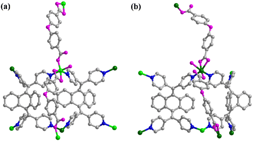
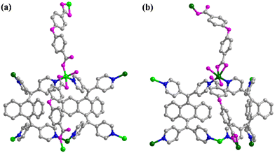
Single-crystal structural analysis demonstrates that 1 belongs to a monoclinic crystal system with a P21/c space group (Table 1) and exhibits a self-interpenetrated 3D (4,4)-connected framework, which is composed of 4-connected Ni2+, 4-connected L units and OBA2− bridges (Fig. 1 and 2). As shown in Fig. S1 (ESI†), the asymmetric unit of 1 contains two Ni2+, one L, two OBA2−, one and a half H2O and three and a half i-PrOH units. The two kinds of crystals are independent Ni2+ with the same coordination environment. Each Ni2+ coordinates with four O atoms from two OBA2− and two N atoms from different L ligands and one water molecule to form a six-coordinated octahedral pattern (Fig. 2). Each L coordinates with two Ni1 and two Ni2 by N atoms from four pyridyl groups (Fig. S2a, ESI†). The dihydroanthracene group is heavily twisted. The angles between two pyridyl groups in one side of dihydroanthracene are 113.833° and 112.056°, which are similar to those from a (116.117° and 111.644°) and b (113.793° and 108.01°).
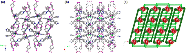 |
| | Fig. 1 Self-interpenetrated 3D framework of 1 along the a-axis (a) and c-axis (b) (Ni1, bright green; Ni2, sea green; N, blue; O, pink; and C, grey). (c) Topology of 1 (Ni1 node, bright green; Ni2 node, sea green; and L node, red). | |
 |
| | Fig. 2 Coordination and connection of Ni1(II) (a) and Ni2(II) (b) (Ni1, bright green; Ni2, sea green; N, blue; O pink; and C, grey). | |
Two OBA2− units show the same coordination and connection manners. Each OBA2− bonds with one Ni2+ by one carbonyl O atom and chelates with one Ni2+ by two O atoms from another side carbonyl (Fig. S2b, ESI†). Therefore, each OBA2− unit is in a μ2-η1η1η1 manner. The lengths of the Ni–N and Ni–O bonds range from 2.047(3) Å to 2.111(3) Å and 2.006(2) Å to 2.207(3) Å, respectively.
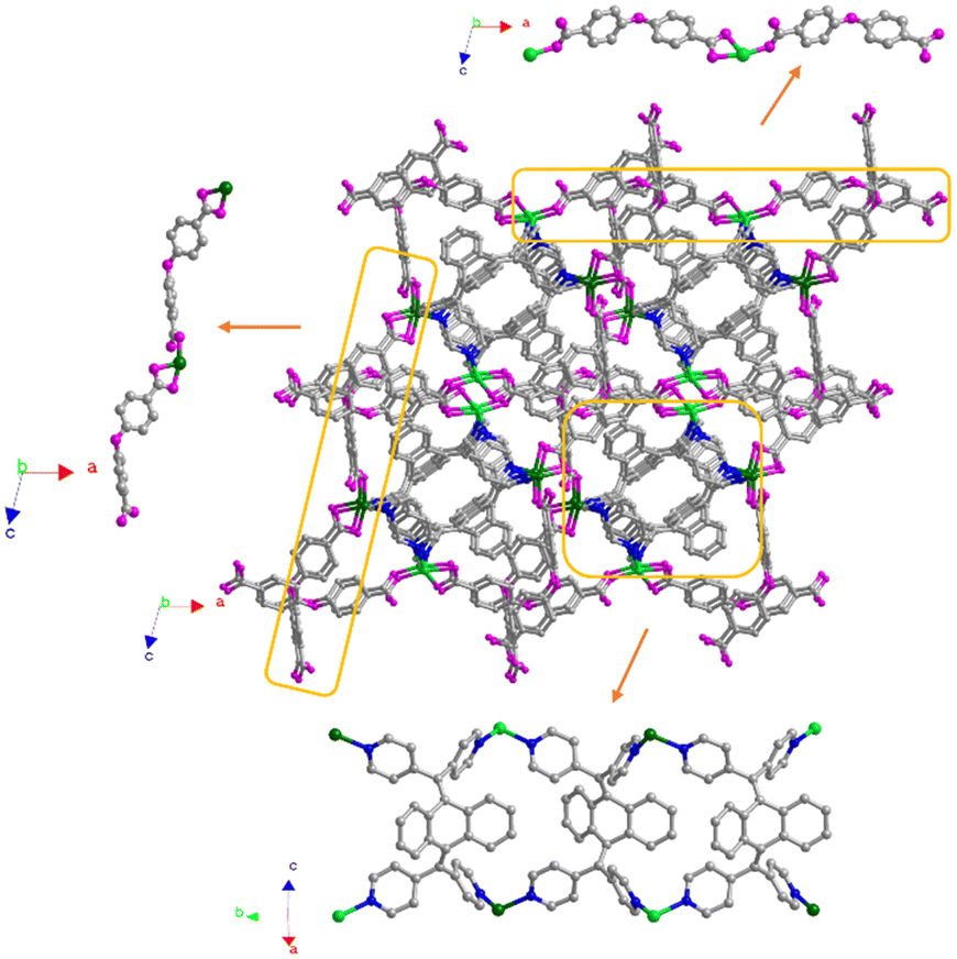
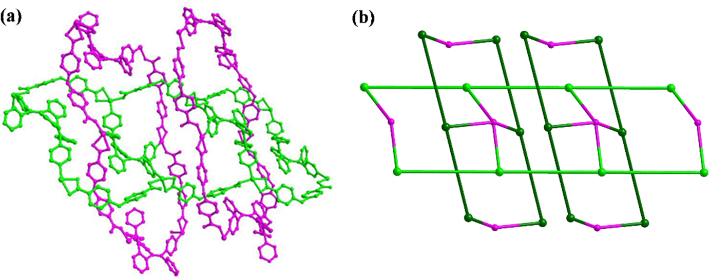
OBA2− bridges connect with Ni1 or Ni2 in a μ2-η1η1η1 manner, which results in 1D chains along the a-axis or c-axis (Fig. 3 and Fig. S3a, b, ESI†). Each L links with two Ni1 and two Ni2 and acts as 4-connected units through its four pyridine groups to form a 1D double-chain architecture along the b-axis (Fig. 3 and Fig. S3c, ESI†). 1D chains fabricated by OBA2− and Ni1/Ni2 connect with 1D double-chains constructed using L, Ni1 and Ni2 units to form a 3D framework (Fig. 3). Interestingly, 1D chains fabricated by OBA2− and Ni1/Ni2 units interpenetrate each other in this 3D framework (Fig. 4).
 |
| | Fig. 3 Formation of the self-interpenetrated 3D framework. | |
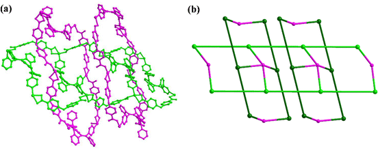 |
| | Fig. 4 Self-interpenetrated structure and topology. | |
L connects with two Ni1 and two Ni2 in a 4-connected manner. Each Ni1/Ni2 links with two L units and another two NI1/Ni2 and is still in a 4-connected manner. From the perspective of topology, L and Ni1/Ni2 units can be regarded as 4-connected nodes. The Schlafli vertex symbols are 42.62.82 and 4.64.8 for L and Ni1/Ni2 nodes, respectively. Therefore, 1 has a binodal (4,4)-connected topology with a Schlafli symbol of (42.62.82)(4.64.8)2 (Fig. 1c). A lot of self-interpenetrated MOFs have been reported;42,43 however, this binodal (4,4)-connected topology with a Schlafli symbol of (42.62.82)(4.64.8)2 is very scarce.44,45
Previously, L was used by our group to construct two MOFs, {[Cd1.5(L)(HOBA)(EtOH)(SO4)(H2O)]·3H2O}n (a)38 and [Cd2L(HBTC)2·2H2O·2i-PrOH]n (b).39 They show a 1D triple chain and a 2D (4,6)-connected network, respectively. However, 1 features a more complicated 3D self-interpenetrated (4,4)-connected structure. L acts as a 4-connected building unit in these MOFs. The involvement of large coordinated molecules (EtOH and SO42−) and rigid HBTC2− in a and b should limit the extension of their structure. 1 has a small H2O coordinated molecule and long and flexible OBA2− bridges, which results in a 3D self-interpenetrated architecture.
PXRD and TGA
The PXRD pattern of 1 shows good agreement with the simulated pattern from its single crystal data, which proves that 1 has a highly pure phase (Fig. S4, ESI†). The PXRD pattern of 1′ can agree well with that of 1 (Fig. S4, ESI†), which reveals that 1′ and 1 are isomorphic. After soaking in water for 7 days, the PXRD pattern and the mass of 1 have almost no changes (Fig. S4 and S5, ESI†), indicating the excellent water stability of 1. 1 was immersed in solutions with different pH values for 24 h. The PXRD patterns of 1 agree well with the original pattern within the pH range of 3–11 (Fig. S6, ESI†). As shown in Fig. S7 (ESI†), in a N2 atmosphere, 1 loses its solvent isopropanol and coordinated water before 159 °C, but the framework can remain unchanged up to 338 °C. Therefore, 1 exhibits excellent water, pH and thermal stabilities.
Photochromism and luminescence of 1
The light green crystals were collected and dried in air (Fig. S8a, ESI†). After being irradiated under UV light, the color of the crystals after soaking in H2O changed from green to yellow (Fig. S8b and c, ESI†). This is an obvious photochromic phenomenon. The suspension of 1 shows excitation and emission at 350 nm and 469 nm, respectively (Fig. 5). The color of the H2O suspension of 1 is colorless. After being irradiated under UV light, the colorless suspension turns its color to yellow. The suspension of 1′ shows a new excitation peak at 400 nm and the corresponding emission peak at 504 nm (Fig. 5). This indicates that the excitation and emission wavelengths are red-shifted. The photochromism from 1 to 1′ should be attributed to photocyclodehydrogenation of L units.38
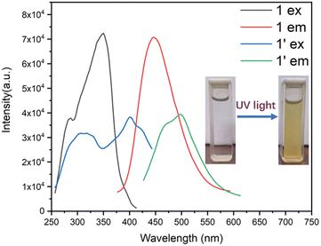 |
| | Fig. 5 Excitation and emission of 1 and 1′. | |
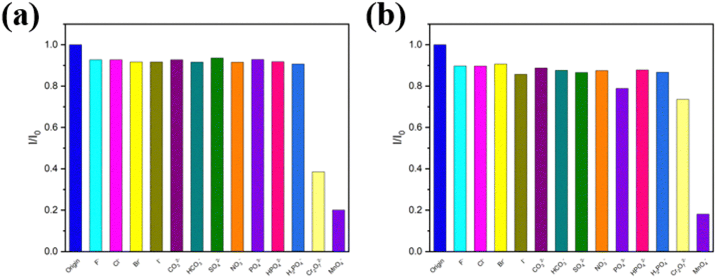
MnO4− sensing
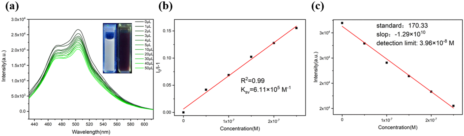
A 50 μL solution with different anions (5 mM) was added into 2 mL H2O suspensions of 1 and 1′. As shown in Fig. 6, MnO4− and Cr2O72− can quench the emission of the suspension of 1. Only MnO4− can quench the emission of the suspension of 1′. Therefore, 1 and 1′ can act as promising MnO4− sensing materials. Titration experiments of 1 and 1′ toward 1 mM and 0.1 mM solutions of MnO4− were carried out, respectively (Fig. 7a and 8a).
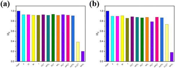 |
| | Fig. 6 Quenching efficiency of 1 (a) and 1′ (b) upon addition of different anions. | |
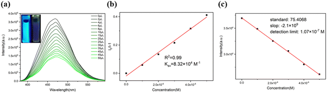 |
| | Fig. 7 (a) Emissions of 1 dispersed in H2O with the addition of MnO4− aqueous solution (1 mM) (λex = 350 nm and λem = 469 nm). (b) S–V plot of 1 for sensing MnO4−. (c) LOD of 1 for sensing MnO4−. | |
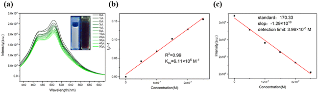 |
| | Fig. 8 (a) Emissions of 1′ dispersed in H2O with the addition of MnO4− aqueous solution (0.1 mM) (λex = 400 nm and λem = 504 nm). (b) S–V plot of 1′ for sensing MnO4−. (c) LOD of 1′ for sensing MnO4−. | |
The quenching efficiency can be quantitatively analyzed using the Stern–Volmer (S–V) equation: I0/I = 1 + Ksv × [M]. The Ksv value of 1 for MnO4− is found to be 8.32 × 10⁴ M−1 (Fig. 7b). In addition, 3σ/k (where σ represents the standard deviation and k represents the slope) is used to calculate the LOD. The LOD value of 1 for detecting MnO4− is calculated as 1.07 × 10⁻⁷ M (Fig. 7c). But the Ksv and LOD values of 1′ for detecting MnO4− are found to be 6.11 × 10⁵ M−1 and 3.96 × 10⁻⁸ M, respectively (Fig. 8b and c). The Ksv value of 1′ is over 7 times higher than that of 1, and the LOD value of 1′ is about 3 times lower than that of 1. More importantly, the LOD value of 1′ reaches the nearly lowest level; the Ksv value of 1′ is the second highest in the reported MOF-based MnO4− sensors (Table 2). Therefore, 1′ has ultrahigh MnO4− sensing sensitivity. The emission intensities and PXRD patterns of 1 (Fig. S9, ESI†) and 1′ (Fig. S10, ESI†) remain basically unchanged after 4 cycles of MnO4− sensing, which reveals their good MnO4− sensing recyclability.
Table 2 Luminescent MOF-based MnO4− sensors in aqueous medium
|
|
K
sv (M−1) |
LOD (mM) |
Ref. |
|
1
|
8.32 × 10⁴ |
1.07 × 10⁻⁴ |
This work |
|
1′
|
6.11 × 10⁵ |
3.96 × 10⁻⁵ |
This work |
| {[Cd1.5(L)(HOBA)(EtOH)(SO4)(H2O)]·3H2O}n |
3.74 × 10⁶ |
4.11 × 10⁻⁵ |
38
|
| [Cd2L(HBTC)2·2H2O·2i-PrOH]n |
4.08 × 10⁵ |
9.33 × 10⁻⁵ |
39
|
| {Zn(L)(TPA)·H2O}n |
1.16 × 10⁵ |
8.0 × 10⁻⁴ |
40
|
| {[Eu2Na(Hpddb)(pddb)2(CH3COO)2]·2.5(DMA)}n |
2.84 × 10³ |
5.99 × 10⁻³ |
46
|
| [Zn(L)2]·2DMF |
1.92 × 10⁴ |
2.34 × 10⁻⁴ |
47
|
| [(H2bpp)·[(UO2)2]-(nip)3]·H2O |
1.88 × 10⁴ |
1.79 × 10⁻³ |
48
|
| {[Ln2(L2)2(H2O)5]·3H2O}n |
8.47 × 10³ |
1.36 × 10⁻³ |
49
|
| In(OH)bpydc (In-MOF) |
2.07 × 10⁵ |
1.47 × 10⁻⁴ |
50
|
| TMU-41(OMS) |
3.2 × 10⁵ |
3 × 10⁻⁵ |
51
|
| [Ba3La0.5(μ3-L)2.5(H2O)3(DMF)] |
7.73 × 10³ |
2.8 × 10⁻⁴ |
52
|
100 μL of 5 mM H2O solution of anions was added to 2 mL of a stable water suspension of 1. These anions, except for Cr2O72−, only slightly quenched the emission of 1. Cr2O72− can quench the emission of 1. When the same volume of H2O solution of MnO4− (1 mM) was added into the above suspensions, the emission of 1 was obviously quenched (Fig. 9a). And for 1′, these anions only slightly quenched the emission of 1′. While the same volume of 0.1 mM MnO4− solution was added into the above suspensions, the emission of 1′ was apparently quenched. So 1′ had excellent MnO4− sensing selectivity (Fig. 9b).
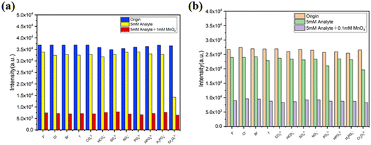 |
| | Fig. 9 Selective MnO4− detection of 1 (a) and 1′ (b) in the presence of different interfering anions. | |
Sensing mechanism
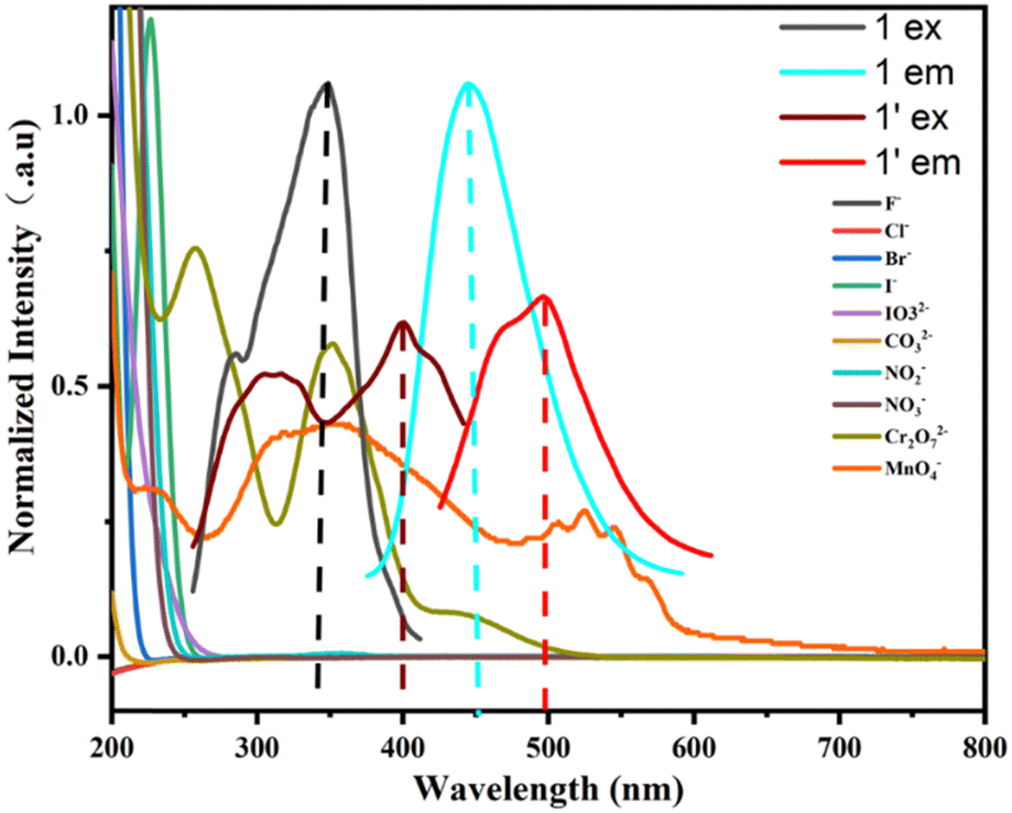
The PXRD patterns of 1 and 1′ are consistent before and after detecting MnO4− (Fig. S4, ESI†), which shows that the frameworks of 1 and 1′ have not collapsed before and after detecting MnO4−. The crystal shapes of 1 and 1′ after detecting MnO4− have no changes (Fig. S7d and e, ESI†). A clear overlap can be observed between the excitation of 1 (260–400 nm) and the absorption of Cr2O72− (320–400 nm) and MnO4− (320–410 nm) (Fig. 10), which leads to competitive energy absorption between Cr2O72−/MnO4− and 1. As a result, both Cr2O72− and MnO4− cause the emission quenching of 1. Accordingly, 1 cannot recognize MnO4− in the presence of Cr2O72−. The new excitation band (350–450 nm) of 1′ overlaps well with the absorption band of MnO4− (320–460 nm) and deviates from the absorption bands of Cr2O72− (320–400 nm) (Fig. 10). Thus, competitive energy absorption can occur only between 1′ and MnO4−. An efficient FRET will occur between 1′ and MnO4− due to a good overlap between the emission of 1′ (420–600 nm) and another absorption band (500–600 nm) of MnO4− (Fig. 10). These cause ultrahigh MnO4− sensing sensitivity and efficient selectivity for 1′.
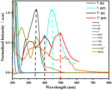 |
| | Fig. 10 UV-vis absorption of anions and excitation and emission of 1 and 1′. | |
Conclusions
An efficient LMOF-based sensor 1 was successfully constructed using a dihydroanthracene-based photochromic organic ligand. 1 exhibits a rare 3D (4,4)-connected self-interpenetrated architecture and has excellent water, pH and thermal stabilities. 1 and 1′ can detect MnO4− in H2O through luminescence quenching effects. Especially, 1′ has extremely high sensing sensitivity and selectivity toward MnO4−. Competitive absorption and FRET should be responsible for the highly efficient MnO4− sensing capability of 1′. This work focuses on the fundamental study of photochromic MOFs including their synthesis, crystal structures, and photochromic, luminescence and sensing properties. This will provide support for the future practical applications of photochromic MOFs such as sensing, anticounterfeiting, etc.
Data availability
Crystallographic data for 1 have been deposited at the CCDC under [2351838] and can be obtained from https://www.ccdc.cam.ac.uk/data_request/cif.
Conflicts of interest
There are no conflicts of interest to declare.
Acknowledgements
This research was supported by the National Natural Science Foundation of China (21671082 and 51432006) and the 111 Project (B13025).
References
- Y. Huang, J. Liang, X. Wang and R. Cao, Chem. Soc. Rev., 2017, 46, 126–157 RSC
 .
.
- H. He, J. Perman, G. Zhu and S. Ma, Small, 2016, 12, 6309–6324 CrossRef CAS PubMed .
- B. Li, M. Chrzanowski, Y. Zhang and S. Ma, Chem. Rev., 2016, 307, 106–129 CAS .
- J. Qin, S. Yuan, C. Lollar, J. Pang, A. Alsalme and H. Zhou, Chem. Commun., 2018, 54, 4231–4249 RSC .
- B. Zhu, Y. Jin, J. Chu, M. Zuo and S. Cui, RSC. Adv., 2022, 12, 6951–6957 RSC .
-
S. Guo, C. Fan, G. Liu and S. Pu, RSC. Adv., 2018, 8, 39854–39864 Search PubMed .
- J. Zhang, S. Ren, H. Xia, W. Jia and C. Zhang, J. Mater. Chem. C, 2020, 8, 1427–1432 RSC .
- A. Mandal, A. Adhikary and A. Sarkar, Inorg. Chem., 2020, 59, 17758–17765 CrossRef CAS PubMed .
- H. Yu, Q. Liu and J. Li, J. Mater. Chem. C, 2021, 9, 562–568 RSC .
- J. Gascon, A. Corma and F. Kapteijn, ACS Catal., 2014, 4, 361–378 CrossRef CAS .
- S. Rogge, A. Bavykina and J. Hajek, Chem. Soc. Rev., 2017, 46, 3134–3184 RSC .
- Z. Wang, X. Yue and Q. Xiang, Coord. Chem. Rev., 2024, 504, 215674 CrossRef CAS .
- S. Ullah, M. Bustam and M. Assiri, Microporous and Mesoporous Mater., 2020, 294, 109844 CrossRef CAS .
- Y. Chen, H. Wu and Y. Yuan, Chem. Eng. J., 2020, 385, 123836 CrossRef CAS .
- H. Daglar, H. Gulbalkan and G. Avci, Angew. Chem., Int. Ed., 2021, 60, 7828–7837 CrossRef CAS PubMed .
- K. Adil, Y. Belmabkhout and R. Pillai, Chem. Soc. Rev., 2017, 46, 3402–3430 RSC .
- J. Li, J. Sculley and H. Zhou, Chem. Rev., 2012, 112, 869–932 CrossRef CAS PubMed .
- P. Horcajada, T. Chalati and C. Serre, Nat. Mater., 2010, 9, 172–178 CrossRef CAS PubMed .
- R. Della, D. Liu and W. Lin, Acc. Chem. Res., 2011, 44, 957–968 CrossRef PubMed .
- P. Horcajada, R. Gref and T. Baati, Chem. Rev., 2012, 112, 1232–1268 CrossRef CAS PubMed .
- C. Martin, K. Park, G. Leith, J. Yu, A. Mathur, G. Wilson, G. Gange, E. Barth, R. Ly, O. Manley, K. Forrester, S. Karakalos, M. Smith, T. Makris, A. Vannucci, D. Peryshkov and N. Shustova, J. Am. Chem. Soc., 2022, 144, 4457–4468 CrossRef CAS PubMed .
- B. Garai, A. Mallick and R. Banerjee, Chem. Sci., 2016, 3, 2195–2200 RSC .
- H. Wentz, G. Skorupskii, A. Bonfim, J. Mancuso, C. Hendon and E. Oriel, Chem. Sci., 2020, 11, 1342–1346 RSC .
- S. Venkateswarlu, A. Reddy, A. Panda, D. Sarkar, Y. Son and M. Yoon, ACS Appl. Nano. Mater., 2020, 3, 3684–3692 CrossRef CAS .
- Y. Xiu and L. Fei, J. Mater. Chem. C, 2015, 3, 22369–22376 Search PubMed .
- L. Wang, J. He and Q. Yang, Environ. Pollut., 2017, 230, 902–910 CrossRef CAS PubMed .
- S. Goldhaber, Regul. Toxicol. Pharmacol., 2003, 38, 232–242 CrossRef CAS PubMed .
- D. Wang, K. Chen, M. Wang, Y. You and X. Zhou, J. Mol. Struct., 2021, 1239, 130486 CrossRef CAS .
- B. Stern, J. Toxicol. Environ. Health, Part A, 2010, 73, 114–127 CrossRef CAS PubMed .
- P. Georgopoulos, A. Roy, M. Yonone-Lioy, R. Opiekun and P. Lioy, J. Toxicol. Environ. Health, Part B, 2001, 4, 341–394 CAS .
- S. Ibrahim and A. Al-Hossainy, J. Mol. Liq., 2020, 318, 114041 CrossRef CAS .
- S. Ibrahim, A. Al-Hossainy, B. Saha and M. El-Aal, J. Mol. Struct., 2022, 1268, 133679 CrossRef CAS .
- Y. Wang, S. Wang and W. Wang, Spectrochim. Acta, Part A, 2020, 229, 117915 CrossRef CAS PubMed .
- J. Ma and W. Liu, Dalton, 2019, 48, 12287–12295 RSC .
- J. Zhang, Q. Qiu and Q. Xiang, Dyes. Pigments, 2021, 196, 109760 CrossRef CAS .
- Z. Hu, B. Deibert and J. Li, Chem. Soc. Rev., 2014, 43, 5815–5840 RSC .
- X. Zhang, F. Chen and Q. Wen, J. Mol. Struct., 2022, 1252, 132183–132190 CrossRef CAS .
- Z. Lin, W. Li, Q. Chen, L. Chen, C. Zhang and J. Zhang, J. Mater. Chem. C, 2022, 10, 1672–1680 RSC .
- J. Zhang, Z. Lin, Y. Yue, Q. Chen, D. Yin and C. Zhang, New J. Chem., 2023, 47, 21986–21993 RSC .
- J. Zhang, L. Chen, Q. Chen, Y. Yue, Q. Chen, D. Yin and C. Zhang, Dyes Pigm., 2023, 220, 111751 CrossRef CAS .
- G. Sheldrick, Acta Crystallogr., Sect. C: Struct. Chem., 2015, 71, 3–8 Search PubMed .
- F. Guo, C. Su, Y. Fan, W. Shi and X. Zhang, Inorg. Chem., 2020, 59, 7135–7142 CrossRef CAS PubMed .
- Y. Qiao, X. Chang, J. Zheng, Z. Chang and M. Yu, Inorg. Chem., 2021, 60, 2749–2755 CrossRef CAS PubMed .
- P. Zhang, B. Li, Y. Zhao, X. Meng and T. Zhang, Chem. Commun., 2011, 47, 7722–7724 RSC .
- D. Zhong, L. Liao, J. Deng, Q. Chen, P. Lian and X. Luo, Chem. Commun., 2014, 50, 15807–15810 RSC .
- B. Li, Q. Yan and G. Yong, J. Mater. Chem. C, 2020, 8, 11786–11795 RSC .
- L. Wang, B. Tu, W. Xu, Y. Fu and Y. Zheng, Inorg. Chem., 2020, 59, 5004–5017 CrossRef CAS PubMed .
- M. Lee, J. Kim and J. Sessler, Chem. Soc. Rev., 2015, 44, 4185–4191 RSC .
- Z. Sun, J. Sun, L. Xi, J. Xie, X. Wang, Y. Ma and L. Li, Cryst. Growth Des., 2020, 20, 5225–5234 CrossRef CAS .
- J. Wu and B. Yan, J. Colloid Interface Sci., 2017, 504, 197–205 CrossRef CAS PubMed .
- N. Abdollahi and A. Morsali, Acta, 2019, 1064, 119–125 CrossRef CAS PubMed .
- B. Ding, S. Liu, Y. Cheng, C. Guo, X. Wu, J. Guo, Y. Liu and Y. Li, Inorg. Chem., 2016, 55, 4391–4402 CrossRef CAS PubMed .
|
| This journal is © The Royal Society of Chemistry and the Centre National de la Recherche Scientifique 2024 |

 *a,
Yinlong
Yue
a,
Xingyu
Tao
a,
Jiarun
Zhang
a,
Dejing
Yin
b and
Chi
Zhang
*ac
*a,
Yinlong
Yue
a,
Xingyu
Tao
a,
Jiarun
Zhang
a,
Dejing
Yin
b and
Chi
Zhang
*ac