DOI:
10.1039/D2CE00452F
(Paper)
CrystEngComm, 2022,
24, 3612-3620
Supramolecular assemblies of Zn(II) complexes with a D–π–A ligand for sensing specific organic molecules†
Received
31st March 2022
, Accepted 15th April 2022
First published on 18th April 2022
Abstract
It is an attractive but challenging proposition to develop effective fluorescent sensors for detecting specific organic compounds. In this study, we designed and synthesized three Zn(II) complexes, namely [Zn(3N3PY)2](NO3)2·3.5CH3OH (1), [Zn(3N3PY)(BIN)]·1.5DMF (2) and [Zn(3N3PY)(BIN)0.5(HCOO)] (3) (H2BIN = 2,2′-biisonicotinic acid, HCOO− = formate), using a D–π–A ligand 4′-(4-(1H-1,2,4-triazol-1-yl)phenyl)-2,2′:6′,2′′-terpyridine (3N3PY) with mononuclear, one-dimensional (1D) chain and dinuclear structures, respectively. The three-dimensional (3D) supramolecular assemblies were achieved by the linkage of the Zn(II) complexes through π–π and hydrogen bonding interactions. The assemblies demonstrated intense luminescence and exhibited an excellent ability for sensing specific organic molecules namely 2,4,6-trinitrophenol (TNP), 4-methylbenzaldehyde (4-MB) and decabromodiphenyl oxide (DBDPO).
Introduction
In order to protect the environment and human security, the detection of toxic and explosive organic molecules has attracted remarkable attention in the past years.1–5 Nitroaromatic compounds (NACs), like 2,4,6-trinitrophenol (TNP) and 4-methylbenzaldehyde (4-MB), and halogenated flame-retardants, like decabromodiphenyl oxide (DBDPO), are valuable chemicals and have been used in fireworks, plasticizers and flame retardants. However, when they are discharged to the environment, it is inevitable that they will cause damage to the soil quality, fresh air and human health.6 Currently, chemical sensors based on luminescence quenching or enhancement have been well developed owing to their sensitivity, selectivity, rapid response, portability and low cost compared to the existing methods and materials, which usually require specific instruments.7–11 The detection of TNP and 4-MB has been intensively studied by designing and synthesizing novel materials such as conjugated polymers, nanomaterials and coordination polymers (CPs) or metal–organic frameworks (MOFs).12–17 However, research on the fluorescence detection of halogenated flame retardants still needs to be supplemented. Among the recent reported luminescent materials, molecules with the D–π–A structure have attracted great attention, attributed to their intramolecular charge-transfer (ICT) property, and have been proved to be valuable for a wide range of applications, such as OLEDs, sensing, anti-counterfeiting and displays.18–22 A series of novel MOFs utilizing D–π–A ligands with tetrazole and terpyridine groups have been reported to realize the modulation of long persistent luminescence (LPL) by adjustment of their dynamic noncovalent coordination structures.21 It is known that azole is a kind of electron-rich group that is suitable to serve as an intramolecular electron-donor, while pyridine is better at acting as an electron acceptor ascribed to its relative electron-deficient conjugation property.23,24 In fact, it would be interesting to apply this kind of D–π–A molecules combining azole and pyridine groups for fluorescent detection. In our previous studies, we successfully synthesized and characterized a series of metal–organic complexes with D–π–A ligands composed of triazole and terpyridine groups and studied their properties, including fluorescent sensing.25–27 However, it is still rare to utilize these attractive fluorescent materials for detecting TNP, 4-MB and DBDPO.
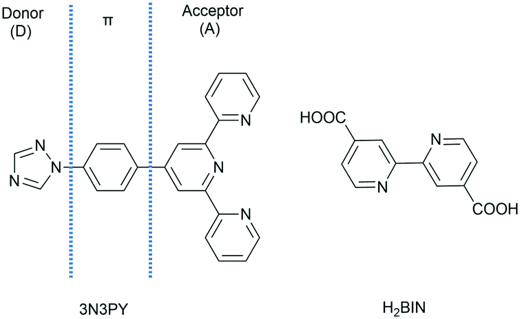
In this work, we designed and synthesized three Zn(II) complexes with a D–π–A ligand of 4′-(4-(1H-1,2,4-triazol-1-yl)phenyl)-2,2′:6′,2′′-terpyridine (3N3PY) together with 2,2′-biisonicotinic acid (H2BIN) serving as structure-directing agents to adjust the stacking mode of the D–π–A molecule of 3N3PY (Scheme 1), and found that they showed high efficiency for the fluorescence detection of TNP, 4-MB and DBDPO.
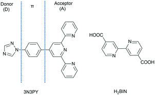 |
| | Scheme 1 Molecular structures of 3N3PY and H2BIN. | |
Experimental
Materials and general methods
All commercially available solvents and chemicals were of reagent grade and were used directly without further purification. The ligand 3N3PY was prepared according to the previously reported method.25–27 Powder X-ray diffraction (PXRD) data were collected on a Bruker D8 Advance X-ray diffractometer with Cu Kα (λ = 1.5418 Å) radiation. Thermogravimetric analysis (TGA) was carried out on a Mettler-Toledo (TGA/DSC1) thermal analyzer with a heating rate of 10 °C min−1 under a N2 atmosphere. FTIR-ATR spectra were measured on a Bruker Tensor II infrared spectrophotometer equipped with a diamond ATR module in the range of 400–4000 cm−1. Elemental analyses for C, H, and N were performed on an Elementar Vario MICRO elemental analyzer. Fluorescence spectra were recorded on a Perkin Elmer LS-55 fluorescence spectrophotometer. Fluorescence lifetimes and the absolute quantum yields (ab-QYs) were measured on a HORIBA Jobin Yvon Fluoromax-4 spectrometer. UV-vis spectral measurements were conducted on a Shimadzu UV3600 spectrophotometer.
Synthesis of [Zn(3N3PY)2](NO3)2·3.5CH3OH (1)
A mixture of 3N3PY (18.82 mg, 0.05 mmol), H2BIN (12.21 mg, 0.05 mmol) and Zn(NO3)2·6H2O (14.87 mg, 0.05 mmol) in methanol (4 mL) was reacted at 110 °C for 72 h under solvothermal conditions. Block crystals were obtained in a 32% yield after cooling down to ambient temperature. Anal. calcd for C49.5H46N14O9.5Zn: C, 56.39; H, 4.40; N, 18.60%. Found: C, 56.39; H, 4.51; N, 18.03%. FTIR-ATR (cm−1, Fig. S1†):![[thin space (1/6-em)]](https://www.rsc.org/images/entities/char_2009.gif) 3408 (s), 3098 (m), 1604 (s), 1525 (s), 1460 (s), 1395 (vs), 1316 (vs), 1146 (w), 1028 (s), 962 (m), 831 (m), 779 (m), 727 (w), 648 (w), 516 (w).
3408 (s), 3098 (m), 1604 (s), 1525 (s), 1460 (s), 1395 (vs), 1316 (vs), 1146 (w), 1028 (s), 962 (m), 831 (m), 779 (m), 727 (w), 648 (w), 516 (w).
Synthesis of [Zn(3N3PY)(BIN)]·1.5DMF (2)
Crystals of 2 were obtained by the same method as that used for the synthesis of 1, except that the solvent was changed to N,N-dimethylformamide (DMF) (4 mL) and sealed in a 10 mL glass vial. Block crystals of 2 were isolated in a 62% yield. Anal. calcd for C39.5H32.5N9.5O5.5Zn: C, 59.78; H, 4.13; N, 16.77%. Found: C, 59.79; H, 4.38; N, 16.64%. FTIR-ATR (cm−1, Fig. S1†):3436 (w), 3072 (w), 1603 (vs), 1551 (s), 1460 (m), 1356 (s), 1391 (vs), 1346 (vs), 1244 (m), 1135 (m), 1013 (w), 971 (w), 849 (w), 783 (m), 671 (m), 549 (w).
Synthesis of [Zn(3N3PY)(BIN)0.5(HCO2)] (3)
The synthesis of 3 was followed by a similar procedure to the above except for altering DMF to a 4 mL mixed solvent of DMF and H2O (v/v, 3:1). Block crystals of 3 were produced in a 56% yield. Anal. calcd for C30H20N7O4Zn: C, 59.27; H, 3.32; N, 16.13%. Found: C, 59.04; H, 3.32; N, 16.20%. FTIR-ATR (cm−1, Fig. S1†):3085 (w), 2812 (w), 1628 (vs), 1551 (s), 1467 (m), 1369 (s), 1330 (s), 1230 (m), 1135 (w), 1069 (w), 1013 (w), 979 (w), 849 (m), 783 (s), 699 (m), 667 (m), 517 (w).
Sensing analytes in solution
For the fluorescent sensing of TNP, 4-MB and DBDPO, the as-synthesized samples were pulverized and sifted. Then the ground powders were dispersed with ultrasonication in N,N-dimethylacetamide (DMA) or H2O (0.5 mg mL−1) to give stock suspensions. All the emission spectra were recorded in the range of 360–560nm and under excitation at 330nm in DMA suspension, while for those in H2O, the data were recorded from 350 to 550 nm. Each test was performed three times to achieve accurate data.
The study of the anti-interference ability for sensing TNP was carried out by experiments involving a solution of NACs (150 μL, 1 mM), which was initially added to a DMA suspension of the assembly (2 mL, 1 mg mL−1), and then 20 μL 1 mM DMA solution of TNP was added each time.
Sensing TNP in the gas phase
Before the fluorescence sensing of TNP in the gas phase, the ground powders were fixed on polymethyl methacrylate (PMMA) film. PMMA acetone solution (2% wt) was prepared and coated 0.5 mL on a quartz sheet in an 8 mm × 10 mm area and then 8 mg testing powders were adhered on to the transparent polymer substrate. The kinetic fluorescent detection was measured by introducing the prepared film in a 10 mL quartz cell that was previously filled with 20 mg TNP and kept for 48 h to obtain a saturated vapour.
Results and discussion
Crystal structure description
Crystal structure of [Zn(3N3PY)2](NO3)2·3.5CH3OH (1).
Single-crystal X-ray diffraction analysis revealed that 1 crystallized in the triclinic and space group of P![[1 with combining macron]](https://www.rsc.org/images/entities/char_0031_0304.gif) (Table 1). It is noteworthy that the H2BIN ligand did not participate in formation of 1, probably owing to the absence of a base in the synthetic reaction. The structure of 1 was a mononuclear Zn(II) complex, in which Zn1 is coordinated by six nitrogen atoms belonging to two terpyridine groups from two different 3N3PY ligands (Fig. 1a). Complex 1 possessed a similar coordination environment and connecting types of [Zn(3N3PY)2]2+ as a previously reported one [Zn(3N3PY)2](NO3)2·H2O.27 However, the torsion angles between the aromatic rings are different (Fig. S2†) due to the different lattice solvent molecules and crystal-packing modes. The Zn–N bond lengths were in the range of 2.071(2)–2.220(3) Å and the N–Zn–N coordination angles ranged from 75.08(9)° to 172.54(9)° (Table S1†). The [Zn(3N3PY)2]2+ units are linked together to form a three-dimensional (3D) supramolecular assembly (Fig. 1d) through π–π interactions between the triazole and terpyridine groups. After calculation, it was determined that there were 41 kinds of π–π interactions in 1 (Fig. S3, Tables S2 and S3†). In addition, there were hydrogen bonding interactions, including C–H⋯N between terpyridine and triazole groups from neighbouring [Zn(3N3PY)2]2+ and C–H⋯O between the 3N3PY and nitrate anions (Fig. 1d, Table S4†) to stabilize the 3D structure.
(Table 1). It is noteworthy that the H2BIN ligand did not participate in formation of 1, probably owing to the absence of a base in the synthetic reaction. The structure of 1 was a mononuclear Zn(II) complex, in which Zn1 is coordinated by six nitrogen atoms belonging to two terpyridine groups from two different 3N3PY ligands (Fig. 1a). Complex 1 possessed a similar coordination environment and connecting types of [Zn(3N3PY)2]2+ as a previously reported one [Zn(3N3PY)2](NO3)2·H2O.27 However, the torsion angles between the aromatic rings are different (Fig. S2†) due to the different lattice solvent molecules and crystal-packing modes. The Zn–N bond lengths were in the range of 2.071(2)–2.220(3) Å and the N–Zn–N coordination angles ranged from 75.08(9)° to 172.54(9)° (Table S1†). The [Zn(3N3PY)2]2+ units are linked together to form a three-dimensional (3D) supramolecular assembly (Fig. 1d) through π–π interactions between the triazole and terpyridine groups. After calculation, it was determined that there were 41 kinds of π–π interactions in 1 (Fig. S3, Tables S2 and S3†). In addition, there were hydrogen bonding interactions, including C–H⋯N between terpyridine and triazole groups from neighbouring [Zn(3N3PY)2]2+ and C–H⋯O between the 3N3PY and nitrate anions (Fig. 1d, Table S4†) to stabilize the 3D structure.
Table 1 Crystal data and structure refinements for 1–3
| Compound |
1
|
2
|
3
|
|
R
1 = Σ||Fo| − |Fc||/Σ|Fo|.
wR2 = |Σw(|Fo|2 − |Fc|2)|/Σ|w(Fo)2|1/2, where w = 1/[σ2(Fo2) + (aP)2 + bP]. P = (Fo2 + 2Fc2)/3.
|
| Formula |
C49.5H46N14O9.5Zn |
C39.5H32.5N9.5O5.5Zn |
C30H20N7O4Zn |
| Formula weight |
1057.72 |
793.61 |
607.90 |
| Temperature (K) |
193(2) |
296(2) |
193 |
| Crystal system |
Triclinic |
Triclinic |
Triclinic |
| Space group |
P |
P |
P |
|
a (Å) |
8.7701(5) |
9.2514(17) |
9.0484(5) |
|
b (Å) |
14.3922(8) |
12.824(2) |
11.1794(7) |
|
c (Å) |
18.8489(10) |
16.624(3) |
12.8727(7) |
|
α (°) |
78.414(3) |
68.187(6) |
80.467(3) |
|
β (°) |
79.587(3) |
82.616(7) |
80.137(2) |
|
γ (°) |
72.876(3) |
80.538(6) |
77.540(3) |
|
V (Å3) |
2208.5(2) |
1801.3(6) |
1241.43(13) |
|
Z
|
2 |
2 |
2 |
|
Dc (g cm−3) |
1.590 |
1.463 |
1.626 |
|
μ (mm−1) |
0.867 |
0.729 |
1.201 |
|
F (000) |
968 |
700 |
622 |
| Data collected |
27320 |
16347 |
14199 |
| Unique reflections |
8099 |
8119 |
4531 |
| Goodness-of-fit |
1.086 |
1.018 |
1.055 |
|
R
1
[I > 2σ(I)] |
0.0570 |
0.0578 |
0.0371 |
| wR2b [I > 2σ(I)] |
0.1656 |
0.1400 |
0.0974 |
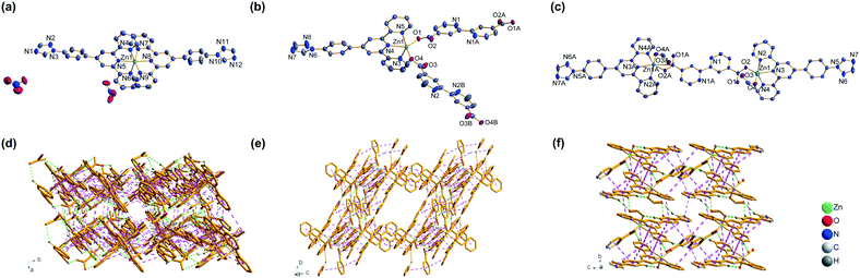 |
| | Fig. 1 Crystal structure of complexes 1 (a), 2 (b) and 3 (c) with ellipsoids drawn at the 50% probability level. Hydrogen atoms are omitted for clarity. Also, 3D supramolecular structure of 1 (d), 2 (e) and 3 (f) constructed with the assistance of π–π interactions (pink lines) and hydrogen bonds (green lines). | |
Crystal structure of [Zn(3N3PY)(BIN)]·1.5DMF (2).
When the reaction was performed in DMF, which can serve as a base, 2 with the deprotonated BIN2− ligand was obtained. As illustrated in Fig. 1b, Zn1 is surrounded by three nitrogen atoms (N3, N4 and N5) from the tridentate N-donor of 3N3PY and two oxygen ones from two different BIN2− with Zn–N bond distances between 2.081(2)–2.234(3) Å and Zn–O ones of 1.962(2) and 1.969(2) Å (Table S1†). In contrast to the mononuclear complex of 1, two [Zn(3N3PY)]2+ units are joined together by one BIN2− ligand to generate a double-arrow subunit [Zn2(3N3PY)2(BIN)]2+ (Fig. S4†), which is further linked together by another BIN2− to produce an infinite one-dimensional (1D) chain structure of 2 (Fig. S5†). Afterwards, the 1D chains pack into a 3D supramolecular structure (Fig. 1e and S6†) with micropores through the noncovalent interactions, including π–π interactions between the 1D chains (Fig. 1e); hydrogen bonding interactions of C–H⋯O (2.43–2.50 Å) between the C–H of 3N3PY and the carbonyl of BIN2− as well as C–H⋯N (2.53 Å) between the C–H of the terpyridine group and the triazole of the ligand 3N3PY (Fig. 1e and Table S4†). The number of π–π interaction types was 23 in 2 (Fig. S3, Tables S2 and S3†). The existence of micropores was tested by the adsorption curve of N2 at 77 K (Fig. S7†).
Crystal structure of [Zn(3N3PY)(BIN)0.5(HCO2)] (3).
Complex 3 was obtained using aqueous DMF as the reaction solvent. As depicted in Fig. 1c, the metal node Zn1 exhibited a similar coordination environment with 2 except for the substitution of a BIN2− by a formate anion resulting from the hydrolyzation of DMF. The formate acted as a terminal ligand in 3, rather than the bridging one of BIN2− in 2, and accordingly, a binuclear structure of 3 was obtained (Fig. 1c). The final 3D supramolecular assembly of 3 (Fig. 1f) is supported by several kinds of weak interactions in 3: (i) π–π interactions between the benzene rings and pyridine groups of 3N3PY (Fig. 1f), where the number of π–π interaction types was 21 for 3 (Fig. S3, Tables S2 and S3†); (ii) hydrogen bonding interactions, including C–H⋯O (2.39–2.58 Å) between the C–H of 3N3PY and the carbonyl group, and the C–H⋯N (2.56 Å) between the C–H bond of terpyridine and nitrogen atom of the pyridine belonging to BIN2− (Fig. 1f, Table S4†).
It is noteworthy that the distinct structures of 1–3 indicated the impact of the reaction medium on the formation of the complexes, since they were prepared using the same reactants with the same stoichiometry of [Zn2+]:[3N3PY]:[H2BIN] = 1:1:1 except for the different solvent. When methanol was used as the reaction solvent in the preparation of 1, no base was present in the reaction mixture to deprotonate H2BIN, resulting in the absence of BIN2− in 1. In fact, the addition of H2BIN was not necessary for obtaining 1. In contrast, DMF could be hydrolyzed to give dimethyl amine and formic acid, which could not only deprotonate H2BIN but also give the formate (COO−)-coordinated complex 3. In addition, 3 could not be obtained by the direct addition of formic acid in the synthesis.
Powder X-ray diffraction (PXRD) and thermogravimetric analysis (TGA).
The consistency between the simulated and experimental PXRD patterns of 1–3 indicated the pure phase of the samples (Fig. S8†). The thermal stability was estimated by TGA under a N2 atmosphere (Fig. S9†). The obtained weight loss of free solvent was 9.89% for 1 and 13.08% for 2, which were consistent with the calculated values of 10.59% for 1 and 13.98% for 2. While for 3, the TG analysis confirmed the absence of free solvent molecules, determined initially by the single-crystal X-ray diffraction analysis. The decomposition temperatures of the assemblies starting from around 280 °C, 300 °C and 350 °C for 1–3, respectively.
Photoluminescence of 1–3.
It has been reported that the D–π–A molecules could display luminescence and consequently, they have been well explored as potential candidates for luminescent materials.18,28 The photoluminescence spectra showed intense emissions at λem = 394 nm for 3N3PY, 409 nm for 1, 411 nm for 2 and 424 nm for 3 under excitation at 330 nm, while almost no fluorescent emission was observed for the ligand H2BIN in the solid state (Fig. S10†). Compared with 3N3PY, obvious red-shifts of the emission maxima were found for 1–3, which may have arisen from the coordination of π-conjugated ligands to Zn(II).29,30 The absolute quantum yields (ab-QYs) of the as-synthesized 1–3 and 3N3PY were respectively measured to be 94.00%, 92.86%, 92.78% and 92.27% under excitation of 330 nm, which may be ascribed to the ICT property of the D–π–A molecule 3N3PY.21
Sensing TNP in solution.
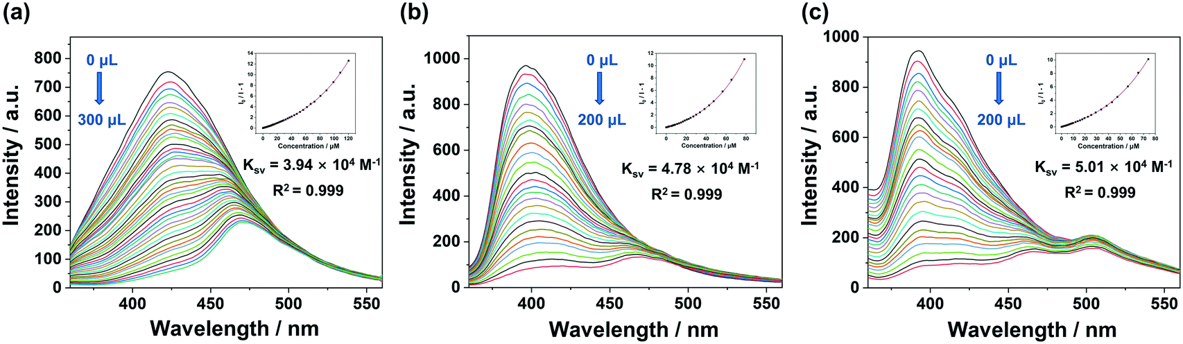
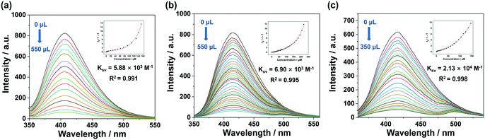
Owing to the excellent luminescent properties of the complexes with the D–π–A molecule 3N3PY, it was considered attractive to explore their abilities for fluorescent sensing. The as-synthesized 1–3 were well ground, sifted and characterized by dynamic light scattering (DLS). The obtained results indicated that the average diameters of the samples were similar and ranged from 6.3 to 8.0 μm (Fig. S11†), which excluded the interference of the size of the samples on the sensing results. PXRD data of as-synthesized and ground crystals were measured at ambient temperature and the characteristic peaks agreed well with the simulated ones generated from single-crystal X-ray diffraction (Fig. S8†), demonstrating the phase purity and maintenance of the structure after grinding. For screening a suitable solvent, 1 was selected as its optimal photoluminescence among the assemblies and the fluorescence sensing ability for TNP was measured in various solvents, including DMA, toluene, water, ethanol (EtOH), DMF, acetonitrile (MeCN) and ethyl acetate (EA) (Fig. S12†). The results illustrated that the best quenching was achieved in the environment of DMA (54.61%) and the detection efficiency was also good when operating in water (42.42%). The fluorescence spectra of 1–3 suspended in H2O and DMA are given in Fig. S13.†
In order to analyse the detection ability of 1–3, fluorescence titration experiments were separately performed in DMA and aqueous suspension, which also reflected the relationship between the fluorescence intensity and the concentration of the test samples. Also, for clearly evaluating the titration results, the fluorescence quenching constant (Ksv) for TNP was calculated by plotting the curves of the relative intensities of I0/I − 1 versus the TNP concentrations (Fig. 2 and 3). The nonlinear Stern–Volmer (S–V) equation (eqn 1) was utilized and the obtained results were satisfactory since the fitted data gave a correlation coefficient (R2) value larger than 0.99:
where
A,
B and
k are constants and
Ksv =
A ×
k.
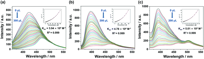 |
| | Fig. 2 Fluorescence titration of 1 (a), 2 (b) and 3 (c) suspended in DMA upon the gradual addition of the DMA solution of TNP. Inset: S–V plot of I0/I − 1 vs. TNP concentration and the calculated quenching constant Ksv. | |
 |
| | Fig. 3 Fluorescence titration of 1 (a), 2 (b) and 3 (c) suspended in water upon the gradual addition of TNP aqueous solution. Inset: S–V plot of I0/I − 1 vs. the TNP concentration and the calculated quenching constant Ksv. | |
The quenching constants Ksv, representing the detection efficiency for TNP, were 3.94 × 10⁴ M−1 for 1, 4.78 × 10⁴ M−1 for 2, 5.01 × 10⁴ M−1 for 3 in DMA suspension, which indicated satisfactory results compared with the reported results (Table S5†). Also, in water detecting medium, the calculated values were 5.88 × 10³ M−1 for 1, 6.90 × 10³ M−1 for 2 and 2.13 × 10⁴ M−1 for 3. The limits of detection (LODs) in DMA suspension for 1–3 were estimated to be 1.71 × 10⁻⁵, 7.32 × 10⁻⁶ and 8.29 × 10⁻⁶ M (Table S6†) and in H2O the corresponding results were 1.17 × 10⁻⁴ M, 7.55 × 10⁻⁵ M and 3.25 × 10⁻⁵ M, respectively (Tables S6 and S7†).
The repeatability of assembly 3 with the best detection performance was examined by detecting TNP in DMA suspension for 5 cycles and the nearly unchanged results indicated its good sensing repeatability (Fig. S14†).
The selectivity of 1–3 for detecting TNP was examined by comparing the quenching efficiency with other NACs upon the addition of the same amount of analyte in DMA suspension of the assembly (Fig. S15†) and it presented remarkable advantages in sensing TNP. A study was performed of the anti-interference ability with this method that further estimated the sensing selectivity, and the data were analyzed by plotting the emission intensity percentage (I/I0) with the added TNP concentration (Fig. S16†). It was clear that the intensity constantly decreased in a stepwise manner, which demonstrated the excellent anti-interference detection ability towards TNP.
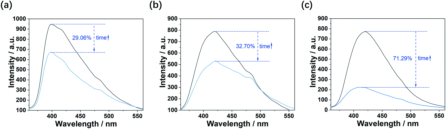
Sensing TNP in the gas phase.
The detection of TNP in the gas phase was performed with the sensors fabricated by fixing the luminescent assembly on PMMA film. Li and co-workers reported a method to detect explosive vapours by luminescent sensors and evaluated the detection performance by analyzing the maximum quenching degree.31 Inspired by these works, the maximum quenching efficiencies for detecting TNP vapour at atmospheric pressure are displayed in Fig. 4 and were found to be in the order of: 1 (29.06%, λem = 420 nm), 2 (32.70%, λem = 428 nm) and 3 (71.29%, λem = 399 nm).
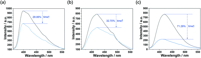 |
| | Fig. 4 Emission spectra (λex = 330 nm) of 1 (a), 2 (b) and 3 (c) films before (black) and after the maximum quenching (blue) by TNP vapour. | |
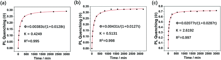
Actually, the analysis results of the detection efficiency were also confirmed to be reliable by comparing the Ksv of different sensors. However, the calculation of Ksv in detecting vapour is inconvenient and generally requires specific sample cells equipped with a pressure transformer to alter their concentration.32,33 Pedrosa and co-workers summarized a practical formula to associate the testing time with the concentration of the analyte vapour, which may be beneficial to calculate Ksv for sensing vapour at atmospheric pressure.34 Accordingly, the modified S–V equation can be written as eqn 2:
| | | Φ = Kk2t/(1 + k2t + Kk2t) | (2) |
where
K =
Q∞ ×
Ksv and
Q∞ is the uptake amount of the analyte vapour at equilibrium. The derivation process of
eqn (2) is presented in the ESI.
†
The kinetic photoluminescent sensing plots for detecting TNP vapour are presented in Fig. 5 and fitted well with the modified S–V equation (eqn (2)) (R2 > 0.99). Given the limited external surface areas and blocked micropores of the fixed fresh samples 1–3, the Q∞ values were considered approximately equal and negligible for comparing their quenching efficiency. As a result, the comparison of Ksv could be reflected by the parameter K and it was found that the calculated K increased in the order of 1 (0.4249), 2 (0.5131) and 3 (2.6192), which is the same trend as for the maximum quenching efficiency (Fig. 5).
 |
| | Fig. 5 Plots for the quenching efficiency (Φ) at 330 nm vs. time under exposure to saturated TNP vapour (black dots) and the fitting curve by the modified S–V model with pseudo-second-order kinetics (red line) for 1 (a), 2 (b) and 3 (c). | |
Sensing 4-MB in solution.
Except for the NACs, metal–organic complexes were also performed well in the recognition of small organic molecules.35 In this work, we investigated the sensing property of 1–3 for 4-MB, benzaldehyde, toluene and acetaldehyde. The detection environment was optimized in DMA suspension considering the quenching efficiency (Fig. S17†), solubility of the analytes, volatility and so on. It can be seen from Fig. S18† that 2 performed well in detecting 4-MB compared to the other probes and analytes. The fluorescence titration experiments for detecting 4-MB by 2 was thus studied for examining the relationship between the alteration of fluorescence and the analyte concentration by the S–V formula and Ksv was calculated to be 1.05 × 10⁵ M−1 (Fig. 6). The LOD of 2 for sensing 4-MB was 6.34 × 10⁻⁶ M (Table S8†).
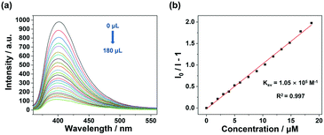 |
| | Fig. 6 Fluorescence titration of 2 (a) suspended in DMA upon the gradual addition of 4-MB solution. (b) S–V plot of I0/I − 1 vs. the 4-MB concentration and the calculated quenching constant Ksv. | |
Sensing DBDPO in solution.
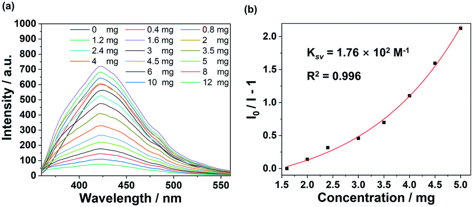
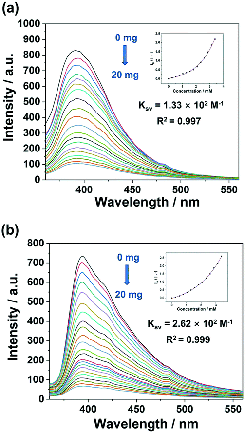
Brominated flame retardants (BFRs) are utilized in a wide variety of commercial products, such as plastics, textiles and electronic/electrical equipment. Due to their toxicity and other adverse factors, the use of certain BFRs, including DBDPO, has been banned or restricted.36 In this work, it is considered meaningful to detect DBDPO in the aqueous environment, since eating a diet including fish and shellfish has been estimated to be the main exposure pathway for BFRs to reach the human body.36 In the detection process with assembly 1, the fluorescence intensity was enhanced initially, which perhaps was induced by the rotation of the conjugated groups in 1,37 and then decreased gradually (Fig. 7). While for 2 and 3, fluorescence quenching occurred directly (Fig. 8). The calculated results for Ksv for the decreasing process were respectively 1.76 × 10² M−1, 1.33 × 10² M−1 and 2.62 × 10² M−1 for 1, 2 and 3 (Fig. 7 and 8). Also, the LODs were separately determined to be 3.91 × 10⁻³ M, 4.86 × 10⁻³ M and 3.01 × 10⁻³ M (Table S9†). As analyzed above, the assemblies 1–3 may provide new probes for detecting BFRs by the fluorescence method.
 |
| | Fig. 7 Fluorescence titration for 1 (a) by suspending in water and accompanied with the gradual addition of DBDPO. (b) S–V plot of I0/I − 1 vs. the DBDPO concentration and the calculated Ksv. | |
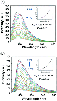 |
| | Fig. 8 Fluorescence quenching titration of 2 (a) and 3 (b) suspended in water upon the gradual addition of DBDPO solid. Inset: S–V plot of I0/I − 1 vs. the DBDPO concentration and the calculated quenching constant Ksv. | |
Possible sensing mechanism.
The mechanism was analyzed by the results from the PXRD, quenching mode, resonance energy transfer (RET), photo-induced electron transfer (PET) experiments and so on. First, no matter in TNP or 4-MB, the crystallinity was almost retained as judged by the PXRD patterns of the assemblies before and after detection, which excluded the variation or collapse of the presented structure (Fig. S19 and S20†) and the crystal structure may have been stabilized by a chelate effect from the tridentate N-donor located at 3N3PY.38 The quenching process was considered to be a static mode for detecting TNP given the rarely changed fluorescence life-time (Table S10, Fig. S21†).39–42 The upward trend of Ksv of 3 > 2 > 1 for sensing TNP is interesting as this is the reverse order of the number of π–π interaction types, i.e.1 > 2 > 3 (Fig. S2†), no matter in DMA or water suspension or detecting TNP vapours, which illustrated the structure–efficiency relationship between the π–π interactions inside these assemblies and their detection performance for TNP. This inferred that the more π–π interactions in the assemblies, the less detection sites for the analyte molecules.43 Furthermore, the possible mechanism of this phenomenon was also analyzed by RET (resonance energy transfer) and photo-induced electron transfer (PET). UV-vis spectroscopy is recognized as a useful method to characterize RET during the detection process. Here, the overlap between the absorption band of TNP and the emission one of 1–3 suggested that energy transfer may occur in the presence of TNP. The wavelength differences between the maximum absorption peak (379 nm) and emission peaks of 1 (422 nm), 2 (396 nm) and 3 (392 nm) were 43, 17 and 13 nm (Fig. S22†), revealing a closer trend and stronger impact of the detection efficiency. As for PET, it is supposed that, during the detection process, the electrons of the assemblies were excited from the valence band (VB) to the conduction band (CB) and then transferred to the LUMO (lowest unoccupied molecular orbitals) followed by returning to the ground state of TNP. Therefore, the PET was analyzed by comparison of the gap energy between the CB of the assemblies and the LUMO of TNP, and it was found that the results decreased in the sequence of 1–3, implying easier electron transfer, which is the same as the order of the quenching efficiency (Table S11 and Fig. S23†). Additionally, the maximum CB was lower than the LUMO of other NACs, such as 4-NCB, with the minimum LUMO among other NACs being −3.342 eV, resulting in a prohibition of the electron transfer and the presence of excellent detection selectivity.3,44,45
Next, as for detecting 4-MB by 2, the detection mechanism was analyzed by comparing the FTIR-ATR spectra of 2 before and after emerging in 4-MB (Fig. S24†). The appearance of the C–H vibration band at 2825 and 2732 cm−1 in the latter curve implied the adsorption of 4-MB in 2, which may be encapsulated in the micropores formed by the noncovalent interactions between the 1D chains (Fig. S6†). As for the quenching process of DBDPO, it probably resulted from the interactions between the bare triazole in the crystals and the electron-deficient benzene ring connected with Br in DBDPO.46
Conclusions
In the present work, three novel Zn(II) coordination complexes were obtained by utilizing the D–π–A molecule 3N3PY to supply intense fluorescence and BIN2− cooperated with a metal node to adjust their stacking structure. To our delight, all of them presented satisfactory efficiency for detecting TNP and performed better with the decrease in π–π interactions in the sensors, no matter in solution or vapour. Additionally, the recognition for 4-MB was also tested and the unique micropores constructed by noncovalent interactions were inferred to be helpful, and so the detection for DBDPO by a fluorescent method was first attempted. This study may provide valuable information for assisting in the design and synthesis of novel efficient fluorescent sensors.
Author contributions
Zi-Qing Huang: conceptualization, investigation, writing – original draft; Jia-Qi Chen: investigation; Shu-Man Zhao: investigation; Zhao-Feng Qiu: investigation; Yue Zhao: formal analysis; Wei-Yin Sun: funding acquisition, supervision; writing – review & editing.
Conflicts of interest
There are no conflicts to declare.
Acknowledgements
We gratefully acknowledge the National Natural Science Foundation of China (grant no. 22171131) and the National Basic Research Program of China (grant no. 2017YFA0303504) for financial support of this work. This work was also supported by a Project Funded by the Priority Academic Program Development of Jiangsu Higher Education Institutions.
Notes and references
- W. P. Lustig, S. Mukherjee, N. D. Rudd, A. V. Desai, J. Li and S. K. Ghosh, Chem. Soc. Rev., 2017, 46, 3242–3285 RSC
 .
.
- S. Xing, Q. Bing, H. Qi, J. Liu, T. Bai, G. Li, Z. Shi, S. Feng and R. Xu, ACS Appl. Mater. Interfaces, 2017, 9, 23828–23835 CrossRef CAS PubMed .
- X. D. Zhang, Y. Zhao, K. Chen, J. H. Guo, P. Wang, H. Wu and W. Y. Sun, Sens. Actuators, B, 2019, 282, 844–853 CrossRef CAS .
- M. E. Germain and M. J. Knapp, Chem. Soc. Rev., 2009, 38, 2543–2555 RSC .
- G. He, H. Peng, T. Liu, M. Yang, Y. Zhang and Y. Fang, J. Mater. Chem., 2009, 19, 7347–7353 RSC .
- T. K. Ghosh, S. Jana and A. Ghosh, Inorg. Chem., 2018, 57, 15216–15228 CrossRef CAS PubMed .
- S. Saglam, A. Uzer, E. Ercag and R. Apak, Anal. Chem., 2018, 90, 7364–7370 CrossRef CAS PubMed .
- D. Shankaran, K. Gobi, K. Matsumoto, T. Imato, K. Toko and N. Miura, Sens. Actuators, B, 2004, 100, 450–454 CrossRef CAS .
- M. Fayazi, M. Ghanei-Motlagh and M. A. Taher, Anal. Methods, 2013, 5, 1474–1480 RSC .
- P. Das and S. K. Mandal, ACS Appl. Mater. Interfaces, 2018, 10, 25360–25371 CrossRef CAS PubMed .
- A. Kalita, S. Hussain, A. H. Malik, U. Barman, N. Goswami and P. K. Iyer, ACS Appl. Mater. Interfaces, 2016, 8, 25326–25336 CrossRef CAS PubMed .
- Y. Salinas, R. Martinez-Manez, M. D. Marcos, F. Sancenon, A. M. Costero, M. Parra and S. Gil, Chem. Soc. Rev., 2012, 41, 1261–1296 RSC .
- F. Akhgari, H. Fattahi and Y. M. Oskoei, Sens. Actuators, B, 2015, 221, 867–878 CrossRef CAS .
- Z. Hu, B. J. Deiber and J. Li, Chem. Soc. Rev., 2014, 43, 5815–5840 RSC .
- X. Zhang, W. Wang, Z. Hu, G. Wang and K. Uvdal, Coord. Chem. Rev., 2015, 284, 206–235 CrossRef CAS .
- X. Y. Zhang, S. M. Zhao, R. Li, Z. H. Xu, M. Y. Wang, Y. F. Jiang, K. Chen, Y. Zhao and W. Y. Sun, Dalton Trans., 2021, 50, 4408–4414 RSC .
- Z. H. Xu, Z. Q. Huang, X. H. Liu, Y. Zhao, Y. Lu and W. Y. Sun, Dalton Trans., 2021, 50, 2183–2191 RSC .
- C. Gao, M. K. Hossain, M. A. Wahab, J. Xiong, B. M. Qiu, H. Luo and W. Li, Dyes Pigm., 2019, 160, 909–914 CrossRef CAS .
- Q. Li, J. Hu, J. Lv, X. Wang, S. Shao, L. Wang, X. Jing and F. Wang, Angew. Chem., Int. Ed., 2020, 59, 20174–20182 CrossRef CAS PubMed .
- B. Zhai, M. K. Hossain, Z. Hu, B. Liu, W. Li and C. Gao, Bull. Korean Chem. Soc., 2019, 40, 1123–1127 CrossRef CAS .
- Z. Wang, C. Y. Zhu, Z. W. Wei, Y. N. Fan and M. Pan, Chem. Mater., 2019, 32, 841–848 CrossRef .
- Z. Wang, C. Y. Zhu, S. Y. Yin, Z. W. Wei, J. H. Zhang, Y. N. Fan, J. J. Jiang, M. Pan and C. Y. Su, Angew. Chem., Int. Ed., 2019, 58, 3481–3485 CrossRef CAS PubMed .
- Z. Gong, R. Wang, Y. Jiang, X. Kong, Y. Lin, Z. Xu, G. Zhou, J. M. Liu, K. Kempa and J. Gao, Org. Electron., 2021, 92, 106102 CrossRef CAS .
- L. Yang, Y. Liao, J. K. Feng and A. M. Ren, J. Phys. Chem. A, 2005, 109, 7764–7774 CrossRef CAS PubMed .
- P. Wang, L. Luo, T. A. Okamura, H. P. Zhou, W. Y. Sun and Y. P. Tian, Polyhedron, 2012, 44, 18–27 CrossRef CAS .
- P. Wang, T. A. Okamura, H. P. Zhou, W. Y. Sun and Y. P. Tian, Chin. Chem. Lett., 2013, 24, 20–22 CrossRef CAS .
- P. Wang, Z. Li, G. C. Lv, H. P. Zhou, C. Hou, W. Y. Sun and Y. P. Tian, Inorg. Chem. Commun., 2012, 18, 87–91 CrossRef CAS .
- D. Zhao, X. H. Liu, Y. Zhao, P. Wang, Y. Liu, M. Azam, S. I. Al-Resayes, Y. Lu and W. Y. Sun, J. Mater. Chem. A, 2017, 5, 15797–15807 RSC .
- J. P. Zou, Q. Peng, Z. H. Wen, G. S. Zeng, Q. J. Xing and G. C. Guo, Cryst. Growth Des., 2010, 10, 2613–2619 CrossRef CAS .
- X. Zheng, L. Zhou, Y. Huang, C. Wang, J. Duan, L. Wen, Z. Tian and D. Li, J. Mater. Chem. A, 2014, 2, 12413–12422 RSC .
- A. Lan, K. Li, H. Wu, D. H. Olson, T. J. Emge, W. Ki, M. Hong and J. Li, Angew. Chem., Int. Ed., 2009, 48, 2334–2338 CrossRef CAS PubMed .
- R. H. Yu, K. Li, Y. Z. Cui, F. R. Tao, B. Zheng, X. S. Ma and T. D. Li, J. Appl. Polym. Sci., 2018, 135, 46708 CrossRef .
- X. Y. Xu and B. Yan, J. Mater. Chem. C, 2016, 4, 8514–8521 RSC .
- F. G. Moscoso, J. Almeida, A. Sousaraei, T. Lopes-Costa, A. M. G. Silva, J. Cabanillas-Gonzalez, L. Cunha-Silva and J. M. Pedrosa, J. Mater. Chem. C, 2020, 8, 3626–3630 RSC .
- Z. Q. Liu, K. Chen, Y. Zhao, Y. S. Kang, X. H. Liu, Q. Y. Lu, M. Azam, S. I. Al-Resayes and W. Y. Sun, Cryst. Growth Des., 2018, 18, 1136–1146 CrossRef CAS .
- L. Trabalon, L. Vilavert, J. L. Domingo, E. Pocurull, F. Borrull and M. Nadal, Food Chem. Toxicol., 2017, 104, 48–56 CrossRef CAS PubMed .
- A. Sussardi, C. L. Hobday, R. J. Marshall, R. S. Forgan, A. C. Jones and S. A. Moggach, Angew. Chem., Int. Ed., 2020, 59, 8118–8122 CrossRef CAS PubMed .
- Z. Q. Huang, Z. H. Xu, X. H. Liu, Y. Zhao, P. Wang and W. Y. Sun, Appl. Organomet. Chem., 2021, 35, e6262 CAS .
-
J. R. Lakowicz, Principles of Fluorescence Spectroscopy, Plenum Press, New York, 2nd edn, 1999 Search PubMed .
- D. Y. Ye, Z. Y. Dong, Y. Q. Pu, G. W. Huang, Y. An and C. W. Lü, Dyes Pigm., 2020, 174, 108016 CrossRef CAS .
- V. Kumar, N. Choudhury, A. Kumar, P. De and S. Satapathi, Opt. Mater., 2020, 100, 109710 CrossRef CAS .
- R. Wang, L. Jiao, X. Zhou, Z. Guo, H. Bian and H. Dai, J. Hazard. Mater., 2021, 412, 125096 CrossRef CAS PubMed .
- S. Kandel, V. Sathish, L. Mathivathanan, A. N. Morozov, A. M. Mebel and R. G. Raptis, New J. Chem., 2019, 43, 7251 RSC .
- N. Seal, A. S. Palakkal, M. Singh, R. Goswami, R. S. Pillai and S. Neogi, ACS Appl. Mater. Interfaces, 2021, 13, 28378–28389 CrossRef CAS PubMed .
- R. Goswami, S. C. Mandal, N. Seal, B. Pathak and S. Neogi, J. Mater. Chem. A, 2019, 7, 19471–19484 RSC .
- H. Xue and J.M. Shreeve, Adv. Mater., 2005, 17, 2142–2146 CrossRef CAS .
|
| This journal is © The Royal Society of Chemistry 2022 |

 a and
Wei-Yin
Sun
a and
Wei-Yin
Sun