
In situ fabrication of silver-based nanostructures using electron beam†
Cuncheng
Ma
a,
Xiangru
Chen
a,
Xiaohua
Tan
a,
Pengfei
Hu
 *a,
Qiang
Li
a,
Yali
Cao
b and
Xue
Liang
*a
*a,
Qiang
Li
a,
Yali
Cao
b and
Xue
Liang
*a
aLaboratory for Microstructures, Shanghai University, Shangda Road 99#, 129 P. O. B, Shanghai 200444, P. R. China. E-mail: hpf-hqx@shu.edu.cn; liangxue@shu.edu.cn; Fax: +86 21 66135030; Tel: +86 21 66135030
bInstitute of Applied Chemistry, Xinjiang University, Urumqi, Xinjiang 830046, P. R. China
First published on 21st March 2018
Abstract
Herein, electron beam irradiation (EBI) was employed to in situ construct Ag-based micro/nanostructures. The composition of AgX@Ag (X = Cl−, Br−, etc.) composites and the size/morphology of silver nanoparticles (Ag-NPs) were tuned by adjusting the electron beam current density (EBCD) and the feeding dose of electrons. A hypothesis of gas–solid fluid-bed composed of atomic gas and nanocrystals is suggested to discuss the nanoparticle constructions with EBI. The low/medium EBCD creates a fixed fluid-bed for AgX@Ag-NPs and piled-pore architecture of Ag-NPs (PPAAg-NPs), while a high EBCD sets up a stirred fluid-bed for monodispersed Ag-NPs. Hopefully, this strategy would promote nanotechnology.
Introduction
Many studies have indicated that the performances of functional materials were affected by the size, shape, composition, and crystallinity of their building units, particularly when their sizes fell into the micro/nanometer size.1–4 Therefore, exploring synthesis strategy aimed at regulating structural parameters for them is and will always be a long-standing goal for scientific and technological development. For example, in 2009, Xia et al. presented a comprehensive review of the shape-controlled synthesis of metal nanocrystals in a solution-based process.5 In that study, the controlled synthesis of silver nanocrystals with various shapes, including the five-fold twinned nanorods, and nanobelts, were selected to elaborate their theme.6–9 Furthermore, several groups designed multiple-step approaches for Ag-based photocatalysts AgnX@Ag-NPs, which mainly involved the ion-exchange reaction for the silver halides or oxysalt of silver, followed by reduction in solution.10–12 Our group has manufactured AgnX@Ag-NPs composites for photocatalyst and silver/silver homojunction assemblies for SERS substrate using a room-temperature mechanochemical technique.13,14EBI has become a powerful tool for engineering the structures of materials and studying the dynamical interface performances of particles at the nanometer or single nanoparticle level, which is of significance both for fundamental research and the potential technological applications.15–19 Wei et al. reported a new EBI phenomenon called local Coulomb explosion to engineer cantilevered multiwalled boron nitride nanotubes.20 José-Yacamán et al. investigated the metal nanoparticle coalescence process with EBI technique and reported that three factors influence the coalescence process: surface diffusion, inherent thermodynamic properties, and coalescence mechanism.21 Latham et al. studied the structural evolution of amorphous Fe oxide nanoparticles from solid spheres into hollow shells via EBI.22 Additionally, in recent years, the EBI methodology is being intensively studied for the construction of the nanoarrays, graphene manipulation, and so on.23–25
In most of the EBI cases, nanoparticles are isolated from the elemental supply source, and the EBI can be considered as an external motivator for the microstructural evolution of nanoparticles. Although EBI can extract the ‘atomic gas’ from the surface of particles as element source for the reconstruction and evolution of nanoparticles,26 the supply of elements will still be scarce and the lack of nanoparticle behavior under irradiation cannot reflect the inherent growth habit. Several groups used AgCl as a precursor, which can provide sufficient elements to construct Ag@AgCl or Ag-NPs by electron beam irradiation. In these cases, Ag+ can be continuously reduced to Ag0 species through EBI, which contributes to the growth of Ag-NPs. The growth of Ag-NPs, the collapse of silver chloride, and the fabrication of Ag@AgCl configuration can be controlled by the current density and the dose of electron beam. Goessens et al.27 investigated the orientation relationship between the AgCl crystal and the Ag particles that are formed on the AgCl crystal by electron beam irradiation. The results indicated that one damaged AgCl region contained an Ag particle with 〈110〉 planes aligned with the 〈100〉 AgCl planes on a (100)Ag(100)AgCl epitaxy. Formo et al.28 investigated the facile synthesis of AgCl:Ag and AgCl mesocubes with edge lengths of up to 500 nm. When these materials were exposed to an electron beam, they morphed into highly textured structures composed solely of silver as a result of the decomposition of AgCl regions in the precursor material. Coupled with experimental observation, Shi et al.29 demonstrated that the growth of s-Ag-NPs was mostly related to the current density of the electron beam used throughout the study of theoretical calculation. In this study, the silver halides, which are sensitive to electron irradiation and can easily be deoxidized to metallic silver, were employed as precursors for controlled fabrication of silver nanostructures and AgX@Ag (AgCl as a delegate) composites by adjusting the electron beam current density (EBCD) and radiation dose. Silver halides can supply the silver elements in situ by reducing silver ions (Ag+) for the growth and evolution of Ag-NPs, providing a possibility for the study of Ag-NP growth. In addition, the gas–solid fluid-bed composed of atomic gas and nanocrystals (FBAg-N) growth mechanism for these micro/nanostructures were suggested and discussed.
Experimental section
AgCl nanoparticles preparation
The AgCl nanoparticles were prepared using a room-temperature solid state reaction method. Briefly, 4 mL polyethylene glycol-400 (PEG-400) was added to 1 mmol silver nitrate (AgNO3) powder and ground in a mortar at room temperature for 0.5 h to obtain a mother batch. Subsequently, 1.2 mmol NaCl was added to the mother batch and ground for 1 h until a brownish-yellow viscous fluid mixture is formed. Then the mixtures were washed with distilled water and ethanol several times. The brown product powder was dried in oven at 60 °C for 4 h and collected for subsequent EBI experiments.EBI process
The EBI experiments of AgCl nanoparticles were carried out on a TEM (JEOL, JEM-2100F). Specimens for TEM were prepared by adding a droplet of alcohol dispersion of AgCl nanoparticles on 230-mesh carbon-coated TEM copper grids, and dried by natural evaporation of alcohol. The construction of Ag-NPs and AgX@Ag-NPs will be investigated by controlling EBCD and dose of electron, which was achieved mainly by adjusting the condenser, spot size, and aperture of the TEM. The details of field emission gun data and EBCD adjustment are stated in Note S1.† Herein, the EBCD value for definition of the irradiation intensity expressions, namely, low, medium, sub-high, and high are set to ≤60, 60–170, 170–240, and ≥240 pA cm−2, respectively. The electron injection dose for AgCl is defined as Q (shown later).Results and discussion
Microstructures of AgCl@Ag-NPs and Ag-NPs and their evolution
Fig. 1 shows the electron microscope (EM) images (containing the transmission electron microscope (TEM), and scanning transmission electron microscope-high angle annular dark field (STEM-HAADF)) of EBI outcomes of AgCl particles with different EBCD and irradiation time (t). It is shown that the medium irradiation (model I: it can be regarded as an electronic shower) can effectively fabricate the AgCl@Ag-NPs composite configuration. These AgCl@Ag-NPs particles displayed fine core–shell hierarchical structures, consisting of large ellipsoidal AgCl core (ca. hundred nanometers orders of magnitude) and their surface loading quasi-spherical Ag-NPs (ca. the size range from several to dozens of nanometers) (Fig. 1a). The EDS-line analysis of a particle indicated the composite structure of Ag@AgCl (Note S2, Fig. S1†). The sizes of Ag-NPs increase with an increase in t under constant EBCD (Fig. 1b). When the Q reaches or exceeds the nAgCl with constant EBCD, AgCl reduces to silver, forming PPAAg-NPs of uniform Ag-NPs (about 100 nm in diameter, Fig. 1c and d). The video of this process clearly shows the collapse of AgCl particles and the formation of Ag-NPs (Note S3† and video-Ag.mp4). Moreover, some adjacent Ag-NPs formed silver/silver (Ag/Ag) homojunction with twinned crystalline structure through sharing of [110] planes (Fig. 1d and e). Moreover, when AgCl was completely reduced by incremental EBCD, the size distribution of Ag-NPs was wider than that obtained in the above case with constant EBCD, in which their diameters ranged from a few to several hundred nanometers (Fig. 1f).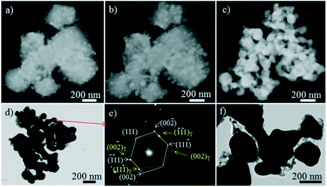 | ||
| Fig. 1 EM images of the objective particles collected after irradiation with different parameters: a and b) STEM-HAADF photographs of AgCl@Ag synthesized under same EBCD (medium, 80 pA cm−2) with irradiation times 15 and 30 seconds, respectively; c and d) STEM-HAADF and TEM pictures of Ag-NPs and their piled pore architecture obtained with sufficient irradiation above EBCD; e) the electron diffraction pattern (EDP) resolution for an Ag/Ag homojunction, displaying twinned crystalline structure; f) TEM images of Ag-NPs constructed with salutatory increasing EBCD. | ||
Different from the abovementioned case, when the beam with high EBCD (model II: it can be called electronic bombing, EBCD > 170 pA cm−2) rushed at AgCl particles, an explosion-type scenario was observed on the stage for Ag-NPs (Fig. 2a). AgCl particles disappeared and were replaced by monodispersed quasi-spherical Ag-NPs (Note S4, Fig. S2†). Furthermore, the sizes of Ag-NPs presented a center-to-peripheral decrease scene from about ten to several nanometers and even less than one nanometer. Simultaneously, some nanoparticles presented multiply twinned particle (MTP) structures, such as five-fold twinned decahedron (five tetrahedrons joined with twin planes rotating on an axis, leaving a gap of 7.35° because of mismatch between the five groups of {111} planes in the tetrahedron (Fig. 2b and c)). In this model, another important trend is that the size of Ag-NPs increases with a decrease in EBCD (comparison of Fig. 2a and d), presenting asymmetric growth profiles with lamellar twinned particle (LTP) structures such as non-symmetric polygons (Fig. 2d–f).
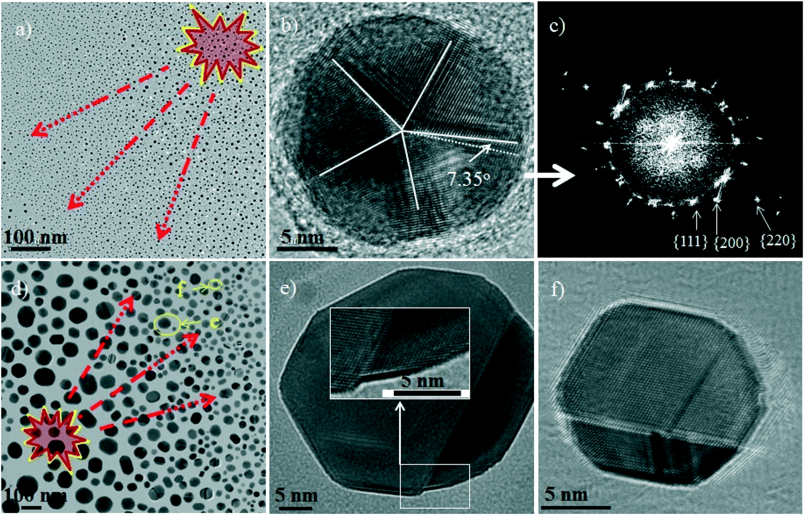 | ||
| Fig. 2 Microstructure resolutions of Ag-NPs prepared with high EBCD: a) TEM images of Ag-NPs harvested with high EBCD (240 pA cm−2); b and c) HRTEM and corresponding fast Fourier transform (FFT) resolution of a decahedron; d–f) TEM and HRTEM images of sample fabricated with sub-high EBCD (180 pA cm−2) showing larger size, twinning structure, and asymmetric growth characteristics. Inset in e) HRTEM image with high magnification of random area of this particle. | ||
Regulation of the composition of AgCl@Ag-NPs (model I) and the microstructure and sizes of Ag-NPs (models I and II)
The regulation of synthesis process with EBI and the structural trends are summarized in a sketch map (Fig. 3). In model I with low/medium EBCD irradiation, the AgCl@Ag composites responded to the Q < nAgCl, while the PPAAg-NPs of Ag-NPs responded to the Q ≥ nAgCl. In model II, the one-step process for the Ag-NPs can be achieved by the bombing-like irradiation with high/sub-high EBCD. In both models, the particle sizes of Ag-NPs were inversely related to the EBCD or proportional to t. In brief, tuning the size and morphology of Ag-NPs in these two models can be achieved by regulating the EBCD or t. Moreover, the asymmetrical growth of Ag-NPs was accompanied by a decrease of EBCD.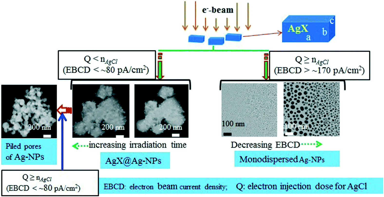 | ||
| Fig. 3 Schematic illustration of the correlation between the structure of outcome and the irradiation parameters in the electron beam irradiation fabrication process. | ||
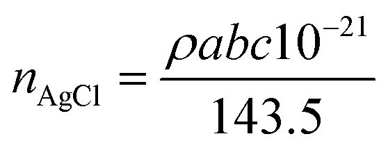
Furthermore, an analysis for adjusting the composition of AgCl@Ag with EBI is roughly discussed as follows. Several approximate boundary conditions are set to discuss this process. First, the ability of AgCl to conduct electrons is considered to be in an off-state and the ejection loss of electrons on the surface of AgCl is neglected. When the electron beam reached the surface of AgCl particles, they will all be in situ requisitioned to reduce the Ag+, but not flown out or ejected. Moreover, AgCl particles are considered to be approximately cuboid. An AgCl cuboid under EBI is schematically shown on the upper right of Fig. 3.
The total amount of AgCl (nAgCl) in a cuboid can be calculated with three-dimensional scale (length a nm, width b nm, and height c nm), density (ρ = 5.56 g cm−3), and molar mass (M = 143.5 g mol−1) as follows:
 | (1) |
The EBCD of the electron beam is set to ID (A cm−2). The surface area of head-on surface of the AgCl particles facing the beam can be calculated as S = a × b. The electron input (Q) is calculated using the following equation:
| Q = ID × S × t | (2) |
It is easy to understand that the output of silver (nAg) is equal to Q. Hence, the total irradiation time (T) for reduction of all the AgCl particles is calculated as follows:
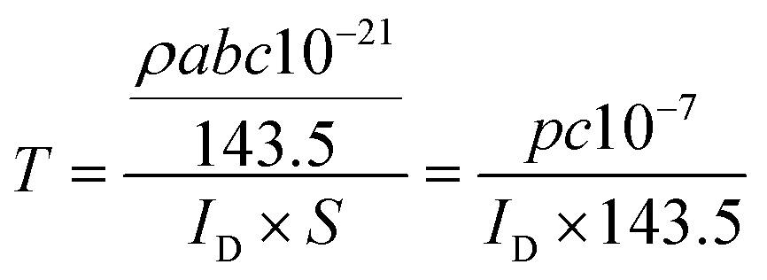 | (3) |
Therefore, the time-dependent molar ratio of the Ag to residual AgCl (nAgCl*) can be deduced by the following equation
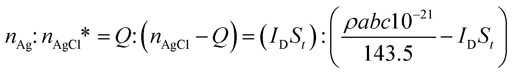 | (4) |
Thus, under a certain current density, the composition of the AgnX@Ag (nAg: nAgnX) can be effectively regulated by monitoring the irradiation time between 0 and T.
Suggestion of atomic gas fluidized bed for building Ag-based nanostructures with EBI
In the present EBI case, the metallic silver is derived from the reduction of Ag+ by electrons, forming the embryonic silver nanocrystals (ENCs-Ag) and atomic gas around them (sketched in Fig. 4). The system can be regarded as a gas–solid fluid-bed, in which the silver atomic gas acts as a fluidizing gas to agitate (mode II) or blow (mode I) the Ag-NPs for fluidization. It should be noted that this fluid-bed is in an open space. The space around them can be regarded as a vacuum (Pspacepressure: 1–4 × 10⁻⁵ Pa) and the pressure of atomic gas is clearly greater than vacuum (Patomicgas > Pspacepressure). Hence, the pressure drop is steep from the center of atomic gas to the outer environment, resulting in the standard deviation of pressure fluctuation (σP) tending to be high and hence, the superficial gas velocity (Ug) cannot be quantitatively measured by a standard orifice flow meter. Moreover, the particle diameter (dP), gas density (ρg), and gas viscosity (μ) varies along with an increase in EBI. Therefore, the minimum fluidization velocity (Umf) and the minimum bubbling velocity (Umb) cannot directly be estimated by the classical method (pressure drop versus Ug) and the Puncochar method (based on the relationship between the Ug and σP), respectively.30–32 Related formulae need to add the correction factor for appropriate use; for example, the Urgun equation for Umf should be modified as Umf = αdp2(ρp − ρg)g/1650 μ (α, correction factor, corresponding simulation study is underway).33 | ||
| Fig. 4 Schematic representation of the fluidized bed composed of nanoparticles and atomic gas. | ||
In model I, the EBI with constant EBCD generated a fixed number of ENCs-Ag and relatively stable flow of silver atomic gas. The system can be regarded as fixed fluid-bed, in which the ENCs-Ag are immobile (the diffusion flow velocity of atomic gas (Ug) is lower than Umf, Ug < Umf), and the atomic gas just flow through the interstices of ENCs-Ag to promote their growth. If the EBCD increases suddenly after producing the first batch of ENCs-Ag, a layout of nanoparticles with large size distribution emerges. Understandably, the spiked enhancement of EBCD will cause a sudden increase of atomic gas. The balance of FBAc-N will be broken to produce a new batch of ENCs-Ag. In the subsequent fluidization process, the sequential batch of ENCs-Ag will grow into mature nanoparticles with different size.
In model II, when the AgX particles were bombed by strong EBI with high EBCD, silver halides were reduced to metallic silver to form a stirred BFAg-N (Umf ≤ Ug, even, Umb ≤ Ug) in an instant (normally, if the silver halide particles are not very large, the following relationship can be satisfied: Q ≥ nAgCl). Therefore, the ENCs-Ag can be transported by fluidizing atomic gas from the center to the periphery. Thus, sputtering scene of particles was observed, in which the diameter of the nanoparticles became increasingly smaller from the center to the periphery due to the center-to-periphery weakening of intensity of atomic gas. In this model, when the EBCD decreased, the low velocity of the silver atom promoted the asymmetric oriented growth of Ag-NPs.
Moreover, it should be noted that the exposed facets of the ENCs-Ag play an important role in intrinsic growth from the crystallographic viewpoint. It is well known that the surface free energy of the planes in the FCC (face center cubic) crystal is of the order of γ{110} > γ{100} > γ{111} based on the calculation for the ‘ideal surface’ model.5 Moreover, the growth energy barriers of the different crystal planes are inversely proportional to their surface free energy. Generally, the chemical reactivity of the (110) facet is greater than that of the (100) and (111) facets, resulting in the silver nanocrystals that easily expose the low-energy (100) and (111) crystalline planes rather than the (110) plane.
Conclusion
In conclusion, electron beam was employed to fabricate the AgX@Ag composites and silver nanostructure with AgX precursor. The composition and microstructure of the object were regulated by tuning the EBCD and the dose of electrons. First, the beam with low/medium EBCD can fabricate AgX@Ag core–shell-shaped architectures and PPAAg-NPs by feeding low or sufficient doses of electrons, respectively. Second, the monodispersed Ag-NPs with center-to-peripheral decrease in size were constructed by irradiation of high-dose and high intensity EBCD. Finally, based on the viewpoint of the ‘ideal surface’ model and quasi-fluidization hypothesis, the formation mechanisms were proposed. The irradiation with low/medium created a fixed fluid-bed for AgX@Ag and PPAAg-NPs, while the high EBCD beam led to stirred fluid-bed for monodispersed small Ag-NPs. The dynamic simulation of atomic cloud fluidized beds in EBI is in progress.Conflicts of interest
There are no conflicts of interest to declare.Acknowledgements
This research was supported by National Nature Science Foundation of China (Grant No. U1503392, 51471101, 21361024, 51472156, and 51472154). Electron microscopy measurements were performed at the analysis and test center of Shanghai University.Notes and references
- J. C. Fraire, L. A. Pérez and E. A. Coronado, J. Phys. Chem. C, 2013, 117, 23090–23107 CAS
.
- M. Banik, P. Z. El-Khoury, A. Nag, A. Rodriguez-Perez, N. Guarrottxena, G. C. Bazan and V. A. Apkarian, ACS Nano, 2012, 6, 10343–10354 CrossRef CAS PubMed
- H. Zheng, R. K. Smith, Y. W. Jun, C. Kisielowski, U. Dahmen and A. P. Alivisatos, Science, 2009, 324, 1309 CrossRef CAS PubMed
- C. Burda, X. Chen, R. Narayanan and M. A. El-Sayed, ChemInform, 2005, 105, 1025–1102 CAS
- Y. Xia, Y. Xiong, B. Lim and S. E. Skrabalak, Angew. Chem., Int. Ed., 2009, 48, 60–103 CrossRef CAS PubMed
- C. Lofton and W. Sigmund, Adv. Funct. Mater., 2005, 15, 1197–1208 CrossRef CAS
- Y. Xiong, A. R. Siekkinen, J. Wang, Y. Yin, M. J. Kim and Y. Xia, J. Mater. Chem., 2007, 17, 2600–2602 RSC
- A. Tao, P. Sinsermsuksakul and P. Yang, Angew. Chem., 2006, 118, 4713–4717 CrossRef
- Y. Song, R. M. Garcia, R. M. Dorin, H. R. Wang, Y. Qiu, E. N. Coker, W. A. Steen, J. E. Miller and J. A. Shelnutt, Nano Lett., 2007, 7, 3650–3655 CrossRef CAS PubMed
- Y. Tang, Z. Jiang, G. Xing, A. Li, P. D. Kanhere, Y. Zhang, T. C. Sum, S. Li, X. Chen and Z. Dong, Adv. Funct. Mater., 2013, 23, 2932–2940 CrossRef CAS
- L. Han, P. Wang, C. Zhu, Y. Zhai and S. Dong, Nanoscale, 2011, 3, 2931–2935 RSC
- P. Wang, B. Huang, X. Qin, X. Zhang, Y. Dai, J. Wei and M. H. Whangbo, Angew. Chem., Int. Ed., 2008, 47, 7931–7933 CrossRef CAS PubMed
- B. Lu, F. Zhan, G. Gong, Y. Cao, Q. Zhen and P. Hu, RSC Adv., 2016, 6, 74662–74669 RSC
- P. Hu, Y. Cao, D. Jia, Q. Li and R. Liu, Sci. Rep., 2014, 4, 4153–4161 CrossRef PubMed
- J. W. Liu, J. Xu, Y. Ni, F. J. Fan, C. L. Zhang and S. H. Yu, ACS Nano, 2012, 6, 4500–4507 CrossRef CAS PubMed
- A. V. Krasheninnikov and K. Nordlund, J. Appl. Phys., 2010, 107, 071301–071370 CrossRef
- A. V. Krasheninnikov and F. Banhart, Nat. Mater., 2007, 6, 723–733 CrossRef CAS PubMed
- J. M. Li, Nanotechnology, 2004, 15, 551–554 CrossRef CAS
- J. M. Li, CrystEngComm, 2017, 19, 32–39 RSC
- X. Wei, D. M. Tang, Q. Chen, Y. Bando and D. Golberg, ACS Nano, 2013, 7, 3491–3497 CrossRef CAS PubMed
- M. José-Yacamán, C. Gutierrez-Wing, M. Miki, D. Q. Yang, K. N. Piyakis and E. Sacher, J. Phys. Chem. B, 2005, 109, 9703–9711 CrossRef PubMed
- A. H. Latham, M. J. Wilson, P. Schiffer and M. E. Williams, J. Am. Chem. Soc., 2006, 128, 12632–12633 CrossRef CAS PubMed
- R. Huang, L. Wang, Q. Zhang, Z. Chen, Z. Li, D. Pan, B. Zhao, M. Wu, C. M. Wu and C. H. Shek, ACS Nano, 2015, 9, 11351–11361 CrossRef CAS PubMed
- J. G. Son, J. B. Chang, K. K. Berggren and C. A. Ross, Nano Lett., 2011, 11, 5079–5084 CrossRef CAS PubMed
- M. Keating, S. Song, G. Wei, D. Graham, Y. Chen and F. Placido, J. Phys. Chem. C, 2014, 118, 4878–4884 CAS
- J. O. Bovin, R. Wallenberg and D. J. Smith, Nature, 1985, 317, 47–49 CrossRef CAS
- C. Goessens, D. Schryvers and J. V. Landuyt, Ultramicroscopy, 1992, 40, 243–244 CrossRef
- E. V. Formo, RSC Adv., 2012, 2, 9359–9361 RSC
- G. H. Shi, Scanning, 2013, 35, 69–74 CrossRef CAS PubMed
- Y. Han, J. J. Wang, X. P. Gu, L. F. Feng and G. H. Hu, Ind. Eng. Chem. Res., 2013, 51, 16482–16487 CrossRef
- C. E. Davies, D. Krouse and A. Carroll, Powder Technol., 2010, 199, 107–110 CrossRef CAS
- M. Punčochář, J. Drahoš, J. Čermák and K. Selucký, Chem. Eng. Commun., 1985, 35, 81–87 CrossRef
- S. Ergun, Chem. Eng. Prog., 1952, 48, 89–94 CAS
Footnote |
| † Electronic supplementary information (ESI) available. See DOI: 10.1039/c7ce02244a |
| This journal is © The Royal Society of Chemistry 2018 |