
A review on recent advances for nucleants and nucleation in protein crystallization
Ren-Bin
Zhou
 a,
Hui-Ling
Cao
*b,
Chen-Yan
Zhang
a and
Da-Chuan
Yin
*a
a,
Hui-Ling
Cao
*b,
Chen-Yan
Zhang
a and
Da-Chuan
Yin
*a
aKey Laboratory for Space Bioscience & Biotechnology, School of Life Sciences, Northwestern Polytechnical University, Xi'an, Shaanxi, PR China. E-mail: yindc@nwpu.edu.cn
bShaanxi Key Laboratory of Ischemic Cardiovascular Disease, Institute of Basic & Translational Medicine, Xi'an Medical University, Xi'an, Shaanxi, PR China. E-mail: caohuiling_jzs@xiyi.edu.cn
First published on 25th January 2017
Abstract
The elucidation of protein structures by X-ray crystallography remains the most effectual method to provide accurate structural details at atomic resolution for rational drug design and other biotechnological research studies. Also, emerging applications of protein crystals as ordered nanostructure scaffolds for catalysis, imaging, and drug delivery are attracting much attention. However, the first step of these applications is obtaining high-quality crystals, which is still an obstacle. Successful crystallization requires two steps: nucleation and crystal growth, while the nucleation is a precondition for harvesting the crystal of interest. So controlling protein nucleation may be an alternative breakthrough for this bottleneck. It is well known that nucleants can induce protein crystallization and improve crystal quality, so investigation on the nucleants that can be universally used for any protein crystallization is ongoing. This manuscript reviews the advances that have been achieved using nucleants in protein crystallization and it is a suitable reference for practical crystallization.
 Ren-Bin Zhou | Ren-Bin Zhou received his bachelor degree in Biomedical Engineering at Northwestern Polytechnical University (Xi'an, China) in 2013. He is currently a Ph.D student at Northwestern Polytechnical University (Xi'an, China). His research interests include protein crystallization and structural biology. |
 Hui-Ling Cao | Hui-Ling Cao obtained her Ph.D in structure biology direction at Northwestern Polytechnical University (Xi'an, China) in 2013. In 2014, she obtained a vice professor position at the Institute of Basic & Translational Medicine, Xi'an Medical University. She also serves as a vice director in Shaanxi Key Laboratory of Ischemic Cardiovascular Disease. She has been researching structure biology and protein crystallization since 2009. Her research interests include protein crystallogenesis, structure biology and drug discovery concerning cardiovascular diseases and cancer. Now she is the author of 30 research papers and presides over five research funds with more than one million RMB. |
 Chen-Yan Zhang | Chen-Yan Zhang, Ph.D, associate professor at Northwestern Polytechnical University, majored in protein crystallization methodology and protein structure biology. |
 Da-Chuan Yin | Da-Chuan Yin obtained his Ph.D in Materials Science at Northwestern Polytechnical University (Xi'an, China) in 1996. He became a lecturer at Northwestern Polytechnical University in 1997. In 2006 he obtained a professor position at the School of Life Sciences, Northwestern Polytechnical University. He has been researching protein crystallization since 2000. Now he is the author of more than 80 scientific research papers and 20 patents. His research interests include protein crystallogenesis, utilization of high magnetic fields for materials processing, and biomedical materials for cell cultures and tissue engineering. |
1. Introduction
In recent decades, there has been increasing interest in understanding the basic mechanisms of biological processes by determining the 3D structures of proteins at the atomic level using X-ray diffraction (XRD).1–4 However, the process of obtaining high-quality crystals for structural determinations is a bottleneck for this method, which delays and often hinders protein crystallography.5–7 Based on the statistics, the likelihood of obtaining high-quality diffraction crystals from purified proteins has been stagnant at approximately 20% for decades.8 The main challenge for protein crystallization is not the inability to obtain protein crystals. What's more disappointing is that the obtained crystals often cannot be used for structural determinations because of their inferior qualities. Multiple advanced methods have been proposed to overcome this obstacle by inducing crystal formation and improving crystal quality. These methods involve controlling the factors that affect protein crystallization (i.e., protein concentration,9 crystallization temperature,10,11 pH value,12 precipitant,13 buffer, additive,14 detergent), utilizing special environments (i.e., microgravity,15 magnetic fields,16,17 electric fields,18 stirring19), developing innovative crystallization methods (microfluidic platforms20), pursuing efficient protein screening kits,21 and so on. All these methods have been proved with varying degrees of success.7 However, the ultimate goal is to characterize a method that is suitable for all protein-to-crystal attempts, where the crystal quality is high enough for diffraction data collection.Nucleants induce protein heterogeneous nucleation and improve crystal diffraction resolution in a controlled manner by providing a substrate to induce nucleation, which subsequently accelerates crystal growth.22 A 1988 study was the first to reveal that nucleation is induced when minerals are used as heterogeneous nucleants.23 It has since been demonstrated that numerous materials (i.e., zeolites, mica and hair) can induce protein nucleation for individual proteins with varying degrees of success, though they cannot be universally used. Investigations on multiple substances as ‘universal’ nucleants for protein nucleation are ongoing. These agents include: (1) mineral substrates for epitaxial nucleation; (2) poor quality crystals or microcrystals for seeding; (3) charged surfaces (i.e., functionalized mica, chemically modified mica, poly-L-lysine surfaces and polymeric film) inducing nucleation by specific interactions; (4) porous nucleants (i.e., porous silicon, bioactive gel-glass, carbon-nanotube-based materials, engineered nano-confined spaces, low density porous or non-porous polystyrene divinylbenzene microspheres (SDB), nanoporous gold nucleants, mesoporous 3D nanotemplates); (5) natural nucleants with their superior biocompatibilities (i.e., minerals, dried seaweed, horse hair, cellulose, hydroxyapatite); (6) the LB nanotemplate (a protein-based nucleant known as the Langmuir–Blodgett homologous protein thin film template) and porous nucleant MIPs (molecularly imprinted polymers). Each nucleant has enabled the attainment of multiple resistant to crystallization crystals and has improved several inferior qualities of the crystals for data collection.
This review starts with a brief assessment of the theoretical issues that surround nucleation and nucleants, including classical nucleation theory, two-step nucleation theory, and the roles and mechanisms of heterogeneous nucleants. Six nucleant types are described based on their detailed mechanisms and specific applications toward protein crystallization. LB nanotemplates and MIPs are novel and effective nucleants and are solely reviewed based on their applications toward inducing nucleation and improving the crystal quality, preparation and mechanism. Lastly, a succinct conclusion is drawn and practice suggestions are provided. This paper is a suitable reference for practical crystallization.
2. Theoretical issues surrounding nucleation and nucleants
2.1 Nucleation theory
An energy barrier must be overcome to form the preliminary crystal nucleus. Crystal nucleation primarily depends on a solution's supersaturation level. Supersaturation, the driving force behind crystallization, signifies that a solution contains the maximum number of dissolved molecules under a given condition at the thermodynamic equilibrium. Because the solid (crystalline) state is more stable than the liquid state (which decreases the Gibbs free energy of the system), supersaturation spontaneously drives the formation and deposition of droplet-like molecular clusters.24,25Gibbs has described the free energy changes that occur within the thermodynamic equilibrium process throughout nucleation. Classical nucleation theory proposes that the free energy of nucleation is associated with two free energies. The negative term is the energy required for bonds to form between molecules in the crystals; this is known as the volume free energy (ΔGv). The positive term corresponds to the unsatisfied bonds that are present on the crystal's surface; this is known as the surface free energy (ΔGs).26 Therefore, the free energy required for nucleation (ΔG) is the sum of the free energy change for the phase transformation (ΔGv) and the free energy change for the surface formation (ΔGs). When nucleation begins, the nuclear volume grows at a minor rate compared to the surface volume till reaching the maximum (ΔG critical) in which the situation changes. Then the nuclear volume grows faster than the nuclear surface volume producing a negative value of the Gibbs free energy, making this process spontaneous. Thus, ΔGv prevails and promotes molecular cluster formation. At the same time, an increase in the free energy of the solid/liquid interface favors dissolution. Therefore, molecular cluster formation depends on the competition between a decrease in ΔGv and an increase in ΔGs, as shown in Fig. 1. The Gibbs free energy for nucleation (ΔG) is a function of the cluster size (r). When it is small, the increasing ΔG promotes dissolution of molecular clusters. As the cluster size increases, ΔG reaches its maximum value and subsequently decreases. This molecular cluster size is known as the critical size. When it is exceeding the critical size, molecular cluster growth becomes energy-favorable and results in nucleation.27
 | ||
| Fig. 1 Free energy diagram for nucleation. Reprinted with permission.27 Copyright 2009, American Chemical Society. | ||
 | ||
| Fig. 2 Crystal nucleation mechanism, classical nucleation theory and two-step nucleation theory. Solid arrows indicate the classical nucleation theory: the ordered clusters occur directly from the supersaturated solution and grow into nuclei. Dashed arrows represent the two-step nucleation theory: disordered protein-rich dense droplet forms, and then crystal nuclei may form inside the droplet. | ||
2.2 Mechanism of promoting nucleation with nucleants
Successful crystallization requires two steps: nucleation and crystal growth. Nucleation is the first major step for crystallization and includes homogeneous and heterogeneous nucleation. Homogeneous nucleation is a random process. Multiple critical nuclei are formed when enough target protein molecules simultaneously assemble in the same region inside the crystallization drop, indicating that homogenous nucleation occurs at higher supersaturation levels. Generally, the energy barrier is low when supersaturation is high, which facilitates the formation of the critical nuclei. However, extremely high supersaturation levels accelerate the crystallization speed, which introduces unfavorable effects, such as structural defects and excessive nucleation, then lead to numerous small crystals instead of a few large ones. Meanwhile, heterogeneous nucleation is often promoted by other substrates and crystallization can be conducted in a controlled manner under metastable conditions.50Nucleants, such as particles or surfaces, can induce heterogeneous nucleation to promote crystallization. To date, a variety of materials have been demonstrated as efficient nucleants, ranging from minerals to porous materials, protein-based and non-protein-based ones, and modified surfaces and natural ones. Generally, it is more advantageous for a protein molecule to be added to an existing nucleus than to form a new nucleus. Also, the pre-existing nucleants lower the energy barrier of the system and stabilize the formed nuclei. In detail, various nucleants may affect heterogeneous nucleation through different mechanisms. For example, minerals as nucleants are for epitaxial nucleation, which requires a match between the crystal lattice of the minerals and that of the protein crystal.23,51 The charged surfaces facilitating nucleation are mainly by electrostatic interactions.52,53 The porous materials can trap protein molecules to form a high local supersaturation to induce nucleation.54 The LB nanotemplate is thought to assist nucleation by causing high electrostatic potential at the film surface, which attracts protein molecules from the solution.55 Meanwhile, a MIP can attract enough protein molecules to overcome the energy barrier for the first step nucleation.56 At the same time, the roughness, topography, microstructure and physicochemical properties of the nucleants affect the heterogeneous nucleation.57
3. Classification and application of nucleants
3.1 Seeding
Seeding is a valuable method for obtaining high-quality crystals of target proteins when regular attempts fail. It is a common practice to transfer several small crystals of the same or different protein as “seeds” under similar or identical crystallization conditions to induce high-quality crystal formation. Seeding can be performed using a cognate protein or by cross-seeding with a different protein.The seeds are visible in macro-seeding, and the seeding procedure is as follows: (1) a suitable single crystal is selected as the seed and collected with a crystal loop from the drop; (2) the crystal is repeatedly washed in a stable mother solution; (3) the washed crystal is transferred to a new crystallization drop for crystallization.61
The seeding procedure for micro-seeding differs from that of macro-seeding and proceeds as follows: (1) multiple small crystals with insufficient qualities in the mother solution are smashed using sonication, by vortexing or with glass rods; (2) the smashed crystals are transferred into a new crystallization drop for crystallization with a seeding needle or micropipette tip. The serial dilution method is used to determine the ideal seeding concentration.62,63
Streak seeding is the easiest and fastest of the three methods. An animal whisker (cat, rabbit or horse hair) is typically used as a seeding wand, which traps the nuclei by it touching the mother crystallization solution.64 A streak line then forms a new drop with the whisker, and the trapped seeds deposit along the streak line.61 Finally, the crystals grow along the streak line, as shown in Fig. 3.
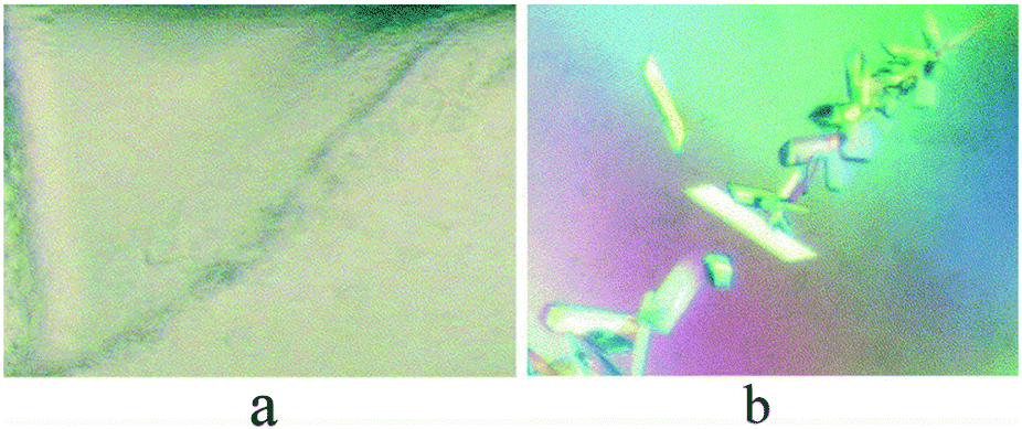 | ||
| Fig. 3 Streak seeding of the mycobacterium tuberculosis RV 2465c protein. (a) Accumulated precipitates along the streak line. (b) Crystals along the streak line. Reprinted with permission.61 Copyright 2003, Elsevier Science (USA). | ||
In summary, the advantages of the seeding methods are obvious. For example, seeding can be utilized to obtain larger and higher diffraction-quality single crystals as well as improve the success rate of protein crystallization,65,66 including membrane proteins.67 Moreover, its facilitation of new crystallization hits is less time-consuming. However, obtaining protein crystals is the precondition for seeding. Recently, automated devices have been developed for seeding trials,63 but they are only employed for primary crystallization screening; crystallization optimization through seeding continues to be manually performed.
3.2 Natural nucleants
Natural nucleants are the preferred heterogeneous nucleants because they have suitable biocompatibilities and are easily obtained. Mineral surfaces comprise the earliest reported natural nucleants, having been described by Alexander Mcpherson and Paul Shlichta in 1988.23 They tested fifty different mineral samples as nucleants for the crystallization of four model proteins—canavalin, concanavalin, catalase B and lysozyme. Their results showed that the mineral substrates induced nucleation and accelerated crystal growth for the four proteins. It is noteworthy that nucleation occurred earlier, at a lower critical supersaturation. Moreover, the mineral substrates altered the crystal habit and unit cell properties. For example, catalase crystals varied from the fine trigonal to the orthorhombic laths. For lysozyme, both the dominant single tetragonal crystals and the fine needle crystals were observed in the presence of the mineral substrates. Regarding unit cell properties, the traditional crystal form of concanavalin B is hexagonal with the space group P63, but a new orthorhombic symmetry P212121 was evident with the mineral substrates.24 The match degree between the mineral substrates and the protein crystal lattices may play an important role in promoting nucleation.51Nine potential natural nucleants have subsequently been tested for the crystallization of ten model proteins in a sparse matrix screen.72 The results showed that four natural nucleants, including dried seaweed, horse hair, cellulose and hydroxyapatite, have positive effects on crystallization. The most efficient natural nucleant was dried seaweed. Predictably, the use of multiple nucleants in the same drop produced the best results. Nonetheless, there were two natural nucleants (fumed silica and carboxymethyl sephadex) that negatively affected crystallization. Positive nucleants for some proteins may act as negative inhibitors for others.
Hair, particularly horse hair and rat whiskers, has been successfully used as a heterogeneous nucleant for many years. It was initially used as a tool for streak seeding; the nucleation-inducing properties of hair itself were discovered by chance. The nucleation-inducing properties of horse hair have been subsequently investigated for three model proteins and a resistant-crystallized recombinant protein, the Fab-D protein. Preliminary investigation demonstrated that horse hair positively affected nucleation at the beginning of the crystallization process.60 Subsequent investigation of human hair fragments as nucleants for the crystallization of three model proteins (lysozyme, glucose isomerase and a polysaccharide-specific Fab fragment) and a resistant-crystallized protein without a 3D structure (the potato serine protease inhibitor) has proved that human hair was a suitable nucleant for obtaining potato protease inhibitor crystals.73
To investigate the mechanistic basis of the hair surface as a nucleant, the following three hair surface properties have been studied: (1) keratins, the most predominant protein in hair; (2) the lipids on the hair surface; and (3) the surface structure and its overlapping terraces. The delipidifying treatment of hair with petroleum ether as the delipidifying agent did not influence crystallization, but the denaturing treatment of hair with ethanol and sodium hydroxide did affect crystallization. Furthermore, an artificial polymer replica for hair did not show a clear preference for nucleation. It is likely that the keratins on the hair surface are essential for crystallization.
3.3 Charged surface
The aggregation of protein molecules is indispensable for nucleation. Protein molecules become charged when they are subjected to specific conditions. This is a useful property because protein molecules can be attracted using an oppositely charged substrate as a nucleant for an electrostatic interaction. In practice, charged surfaces are designed as nucleants to promote protein crystallization. For example, functionalized mica with negatively charged sulfonated polystyrene films and positively charged silanized sheets were prepared as charged nucleants to promote the crystallization of insulin and ribonuclease A.74 As expected, the charged surface shortened the crystallization time and minimized protein consumption. The interactions between the charged surface and the protein molecules enrich the protein molecules around the charged surface to reach local supersaturation, which favors nucleation and crystal growth.43,75 Additionally, the weak force of the charged surface stabilizes the formed nuclei.The fluorinated layered silicate surface has been used as a crystallization nucleant; it promoted lysozyme nucleation while silicates without fluorine suppressed nucleation.76 Nucleation promotion was also observed when fluorine was replaced with hydroxyl groups. The fluorinated layered silicate surface has also been used to crystallize twelve proteins with a wide range of pIs and molecular weights; the negatively charged surface promoted protein nucleation regardless of the net charges and molecular weights of the proteins. The result of the initial screening trials with fluoro-substituted saponite has verified the beneficial effects, which included increasing the initial screening success rate, reducing the nucleation energy barrier and minimizing protein consumption. The mechanism may involve the interaction between the silicate's negatively charged fluorine atoms or hydroxyl groups and the positively charged residues of the protein molecules, which attracts protein molecules causing them to gather at the silicate surface where they form a high local supersaturation zone to form the initial critical nuclei.
Furthermore, chemically modified mica, poly-L-lysine (PLL) surfaces, polymeric films and patterned silicon have been reported as potential nucleants. PLL surfaces are positively charged surfaces that are coated with cationic polymerized lysine. They attract negatively charged molecules and repel the positive ones. The net charge of the lysozyme protein is also positive in the optimum experimental conditions. Because of the repulsive interaction, the PLL surface orients the single lysozyme crystals and diminishes the nucleation number, as shown in Fig. 4.77 However, when negatively charged proteins were used, the PLL surface oriented the single crystals, but it did not affect the nucleation number for the tested proteins.78
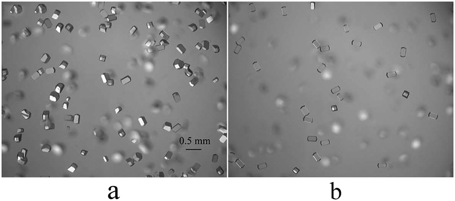 | ||
| Fig. 4 The orientation and reduction of lysozyme crystals. (a) Crystals obtained on normal glass at lysozyme 30 mg ml−1, NaCl 40 mg ml−1 and pH 4.5. (b) Crystals obtained on PLL-coated glass under the same conditions. Reprinted with permission.77 Copyright 2002, Elsevier Science. | ||
Additionally, other charged polymeric surfaces (i.e., sulfonated polystyrene, cross-linked gelatin films with adsorbed poly-lysine, and silk fibroin with entrapped poly-L-lysine or poly-L-aspartate) have been used as heterogeneous nucleants to crystallize concanavalin A and lysozyme. Compared with the siliconized cover glass and the charged polymeric surfaces, the surfaces reduced both the induction time and the necessary protein concentration for nucleation while increased the nucleation density to promote crystallization.79
3.4 Porous nucleants
New porous nucleants have since been developed and successfully proven for promoting protein crystallization. In 2006, mesoporous bioactive gel-glass, which contained a disordered porous medium with a pore size distribution of 2–10 nm, was developed for protein crystallization and became one of the most successful heterogeneous nucleants. The bio-glass promoted the nucleation of various proteins, including multiple resistant-crystallized proteins. The use of the bio-glass was more convenient than using porous silicon because it was easy to add the bio-glass into the crystallization drops.43 The bio-glass has been commercialized as ‘Naomi's Nucleant’ by Molecular Dimensions.
Although bio-glass nucleants induced the crystallization of numerous proteins, its pore sizes and surface chemistry were not easily controlled. Thus, a carbon-nanotube-based material with a controlled pore size and surface chemistry was developed as an efficient nucleant in 2009, as shown in Fig. 5.
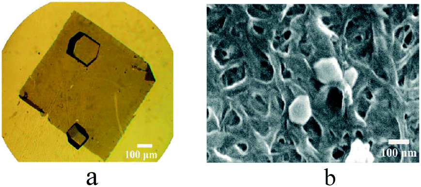 | ||
| Fig. 5 (a) Optical microscopy image of lysozyme crystals on a sheet of transparent buckypaper (films of entangled carbon nanotubes). (b) SEM image of a lysozyme crystal too small to be visible via optical microscopy. Reprinted with permission.81 Copyright 2009, American Chemical Society. | ||
Another study investigated the effects of engineered nano-confined spaces on protein crystallization, which had narrow pore size distributions (pore diameters of 3–4 nm, 6–8 nm, 10–12 nm, 13–15 nm and 17–21 nm) and highly ordered pore structures. Proteins with a wide range of molecular weights, from 14 kDa to 450 kDa, were utilized for the crystallization trials.82 The results were as follows: the 3–4 nm pore diameter worked well for the 12–24 kDa protein crystallization, the 6–8 nm pore diameter was ideal for the 67 kDa protein crystallization, and the proteins with molecular weights of 106–232 kDa were successfully crystallized using the 10–12 nm pore size. The higher molecular weight protein (450 kDa) required a higher 17–21 nm pore size to crystallize. Thus, a wide range of pore sizes would broaden the ability to crystallize different proteins. Surprisingly, human serum albumin (HSA) and concanavalin A-type IV did not crystallize on the porous nucleants with broad pore size distributions,80 while the narrow range pore size ones succeeded (HSA with the 3–4 nm pore diameter; concanavalin A with the 10–12 nm pore diameter). Additionally, the nucleants with slightly larger pore sizes than the gyration diameters of the proteins induced and stabilized the nuclei.82
Nanoporous gold nucleants have also been recently used as heterogeneous nucleants for protein crystallization. When the pore diameter of the nucleant was similar to the diameter of the protein molecule, nucleation on the nucleant surface may have occurred under supersaturation conditions that were milder than usual, which would result in a lower energy barrier for nucleation and higher quality crystals. The interaction between the surface of the dealloying nanoporous nucleant and the target protein molecules played the dominant role in inducing nucleation, rather than the entropy contributions to the free energy of crystallization.83
Low density porous or non-porous polystyrene divinylbenzene microspheres (SDB) are appropriate agents for simultaneously improving nucleation and crystal quality. SDB adsorbs protein molecules at high concentrations and desorbs protein molecules at low concentrations. Thus, the adsorption and desorption properties of SDB enable the formation of relatively high and relatively low concentration regions in the same solution; the high concentration region is favorable to nucleation. Up to a specific size, the nuclei settle in the low concentration region to grow high-quality crystals.84
Mesoporous 3D nanotemplates are also new and efficient heterogeneous nucleating surfaces for protein crystallization. One study became the first to show that concanavalin A and catalase produced different crystal habits under identical crystallization conditions when 3D nanotemplates were used as nucleants. However, the crystals with different crystal habits exhibited the same space group by XRD. The habit modification was the combined result of the porosity and chemistry properties of the 3D nanotemplate.85
The mechanism of nucleation by porous materials is likely explained by four properties. (1) The nanopores trap the protein molecules by diffusion and capillary action, and the protein molecules are restrained inside these pores, which results in a higher local supersaturation and nucleation. The critical nuclei attract the protein molecules causing them to gather and ultimately grow into crystals.86,87 (2) The pore size plays a dominant role in the nucleation rate. Successful nucleation involves two successive processes: the nucleation of the pore filling and nucleation outside the pore under the bulk (homogeneous) conditions. The aforementioned computer simulation shows that the smaller pore size results in a slower nucleation rate outside the pore, and a larger pore size leads to a slower initial pore filling. Therefore, the pore size should be approximately equal to that of the critical nuclei to maximize the nucleation rate.86 (3) The surface chemistry of the pore also contributes to crystal nucleation. The pore surface exhibits a weak chemical attraction and a subsequent weak interaction with the protein molecule to promote nucleation. The attractive interaction facilitates the formation of the high density phase, which is essential for nucleation in the two-step nucleation mechanism. The high density phase formation has been observed and characterized using the light-scattering technique.88 Additionally, the attractive interaction stabilizes the half-formed nuclei.82 (4) The pore traps and immobilizes the protein molecules within, and this lowers the free energy barrier for nucleation.82 Therefore, the nanoscale pore improves the solution's thermodynamic stability, which is favorable for protein folding and nucleation. This was verified in 2006 with a molecular dynamics simulation and experiment.89
3.5 Langmuir–Blodgett thin film template
Surprisingly, the LB nanotemplate successfully crystallized proteins whose structures had not previously been resolved, including bovine cytochrome P450 scc93 and human protein kinase CKII alpha subunit.94 Thus, the novelty and effectiveness of the LB nanotemplate method have generated increasing interest, which is evidenced by its prolific development.87,92,93,95–98
Multiple advanced techniques have been employed to evaluate the LB nanotemplate, including atomic force microscopy,99 Raman spectroscopy,100 small-angle X-ray scattering,101 computer simulation, molecular dynamics,102 Fourier transform infrared (FTIR) spectroscopy, and circular dichroism.99 Lysozyme exhibited a surprising thermal stability during the preparation of the LB nanotemplate, retaining its secondary structure until 200 °C; above 200 °C, the protein aggregated.99 The LB nanotemplate promoted disulfide bond formation (at the S6–S127/S30–S115 position) and hydrogen bond formation in lysozyme, which makes stable crystals easy to obtain.100 Additionally, crystals that were obtained using the LB nanotemplate were highly resistant to radiation damage.103
 | ||
| Fig. 6 Schematic of preparing the LB nanotemplate. | ||
3.6 Molecularly imprinted polymers (MIPs)
In 2011, Naomi E. Chayen became the first to develop a water-based MIP (HydroMIP) as a non-protein nucleant for protein crystallization.120 It is also known as a “smart material” because the MIPs are effective toward increasing the crystal hit number and improving crystal quality.56 Seven proteins—lysozyme, trypsin, catalase, hemoglobin, intracellular xylanase IXT6-R217W, alpha crustacyanin, and human macrophage migration inhibitory factor (MIF)—have been used to evaluate the nucleation-inducing properties of the MIPs. Compared with the non-imprinted polymers and the control without polymers, MIPs induced protein nucleation through their cognate MIP and through other non-cognate MIPs of similar sizes in metastable conditions. Additionally, the diffraction qualities of the crystals that were obtained from the MIPs were superior to that of the control. For example, the complex HIV protein crystals showed diffraction up to 4.2 Å in the presence of lysozyme–MIP, but traditional methods failed to show crystal diffraction beyond 9 Å. Furthermore, the crystals appeared more quickly in the drops that contained MIPs above metastable conditions. Hemoglobin crystals have been observed after 5 days with 22.5% (wt/vol) PEG 3350 that contained Hemoglobin–MIP. At 25% (wt/vol) PEG 3350, all drops formed crystals, but they appeared after 7 days.120,121
To investigate MIP applications to initial screening experiments, four proteins have been screened with and without their cognate MIPs using the popular index screening kit. The useful crystals for two proteins, alpha crustacyanin and intracellular xylanase IXT6-R217W, have not been previously produced. The third protein, human macrophage migration inhibitory factor (MIF), required better resolution. An easily crystallized model protein, trypsin was used as the control.120 The results showed that 8–10% of the screening trials of the aforementioned proteins produced hits, but these hits were missing in the absence of the MIPs. When the concentrations for the MIF and alpha crustacyanin were raised to 15–30%, the prior hits yielded crystals in the absence of MIPs. Thus, MIPs can screen new hits and decrease target protein consumption. Because MIPS are efficient and reproducible, they have been automated for applications in high-throughput robot-based screening trials.122 Moreover, MIPs of differing molecular weights are commercially available for general use.
The MIP method has recently been improved by Chen's group.123 The traditional precipitants were immobilized into MIPs to aid protein crystallization, and high-quality crystals of the flexible N-terminus of the human fragile X mental retardation protein were successfully obtained. The diffraction resolution was improved from 10 Å, which was typical of the traditional methods, to 3 Å when precipitant-immobilized MIPs were present.
Additionally, zwitterionic additives have been immobilized into MIPs to facilitate crystallization. Fortunately, the zwitterion-immobilized MIPs facilitated the formation of higher quality crystals in a shorter time compared with regular MIPs and traditional crystallization trials. The high-quality single crystals of a flexible protein, concanavalin A, have been successfully obtained using this approach.124
Semiliquid MIPs are prepared using a procedure that differs from that of the solid nucleants, as shown in Fig. 7. (1) A functional monomer (AA) and cross-linker (N,N0-methylenebisacrylamide) are dissolved in deionized water. The template protein solution is then added to create the pre-MIP solution. (2) Ammonium persulphate (APS) and N,N,N,N-tetramethylethyldiamine (TEMED) are added to polymerize the solution at room temperature. (3) After polymerization, the formed gels are crushed. (4) The template protein molecule is eluted, and the complementary cavities remain. When the template proteins are introduced into a crystallization trial, they can be easily memorized and rebind to the cavities. The prepared MIPs are stored at 4 °C.120
 | ||
| Fig. 7 Preparation of MIPs. Functional monomer (acrylamide) and template protein were polymerized by a cross-linker (N,N0-methylenebisacrylamide). Then the template protein was removed by elution and protein specific cavities were left. Finally the target protein can be memorized by the complementary cavity and rebind to it to induce crystallization. | ||
4. Concluding remarks
4.1 Conclusion
(1) Seeding is a valuable way to obtain the high-quality crystals of target proteins when regular attempts fail.(2) Natural nucleants, including minerals, dried seaweed, animal/human hair, cellulose and hydroxyapatite, are the preferred heterogeneous nucleants because they have suitable biocompatibilities and are easily obtained.
(3) Charged surfaces (i.e., functionalized mica, chemically modified mica, poly-L-lysine surfaces, polymeric films) can stimulate protein molecule aggregation through their electrostatic interactions to promote protein nucleation and crystallization.
(4) The nanopores of porous nucleants can trap protein molecules to form a high local supersaturation to induce nucleation. Porous nucleants include porous silicon, bioactive gel-glass, carbon-nanotube-based materials, engineered nano-confined spaces, low-density porous or non-porous polystyrene divinylbenzene microspheres (SDB), nanoporous gold nucleants and mesoporous 3D nanotemplates. Bioactive gel-glass is a highly successful heterogeneous nucleant.
(5) The LB nanotemplate is a homologous protein thin film that is made using the Langmuir–Blodgett technology and is transferred to a cover glass to modify the traditional hanging drop vapor diffusion method to promote crystallization.
(6) The complementary cavities in MIPs can induce protein molecules to migrate toward the MIP surface and form a protein-rich phase to overcome the nucleation energy barrier.
4.2 Practical suggestions
To make full use of these nucleants, several practical suggestions are provided below. When crystals are obtained with qualities that are unsuitable for data collection, seeding is the ideal approach. When crystals cannot be obtained by trial and error, it is worth testing effective nucleants to increase the chances of inducing nucleation. The natural nucleants, which have better biocompatibilities and are easily obtained, increase successful crystallization outcomes. Specifically, a nucleant comprised of animal or human hair is the better choice for heterogeneous nucleation. Various porous nucleants are also made suitable by their effectiveness and convenience. They include porous silicon, bioactive gel-glass, carbon-nanotube-based materials, engineered nano-confined spaces, nanoporous gold nucleants and mesoporous 3D nanotemplates. Of the porous nucleants, bioactive gel-glass may be the better choice. Finally, universal nucleants, such as MIPs or LB nanotemplates, are also worth trying to increase the chances of obtaining crystals.5. Future perspectives
Although these nucleants have been demonstrated to successfully aid protein crystallization, they are not so perfect and convenient for protein crystallization. For example, many nucleants only work with some certain cases not suitable for “general” use. The reported “universal” nucleants (LB template thin film and MIPs) also have some drawbacks. A special device is needed to fabricate the LB template thin film and can't be easily obtained in all the structural biology laboratories. Although the MIPs can conveniently yield and have been commercialized, effectively assist the crystallization of globular proteins, protein complexes and membrane proteins, it should notice that the MIPs nucleants can also facilitate salt crystallization when the crystallization buffer contains substantial levels of salt. So an important novel conclusion is to integrate all the nucleants' advantages (effects of epitaxy, special interactions and porous materials nucleation-promoting properties) into the design of more effective nucleants.In the future, it is essential to achieve optimal universality of the heterogeneous nucleants for promoting any protein crystallization, with high automation to dispense them into the crystallization solution for high-throughput screen. Alternatively, the best strategy is to design functional crystallization plates for protein crystallization instead of introducing another heterogeneous nucleant.
Another aspect of protein nucleants that should be addressed is the theory explanation. This is possible to achieve by direct imaging. Scanning electron microscopy, atomic force microscopy and confocal fluorescence microscopy can be employed for this purpose. At the same time, computer simulation is a powerful tool to provide detailed information on nucleants aiding protein crystallization.
Competing interests
The authors have declared no competing interest.Acknowledgements
This work was supported by the National Natural Science Foundation of China (Grant No. U1632126), the National Natural Science Foundation of China (Grant No. 11202167), the Fundamental Research Funds for the Central Universities (Grant No. 3102016ZY039), the Natural Science Foundation of Shaanxi Province, China (Grant No. 2016JM3012), and the Talents Program of Xi'an Medical University (No. 2015 RCYJ01).Notes and references
- H. P. Zheng, K. B. Handing, M. D. Zimmerman, I. G. Shabalin, S. C. Almo and W. Minor, Expert Opin. Drug Discovery, 2015, 10, 975–989 CrossRef PubMed
.
- R. Giege, FEBS J., 2013, 280, 6456–6497 CrossRef CAS PubMed
- A. R. Pearson and A. Mozzarelli, Biochim. Biophys. Acta, Proteins Proteomics, 2011, 1814, 731–733 CrossRef CAS PubMed
- J. L. Grey and D. H. Thompson, Expert Opin. Drug Discovery, 2010, 5, 1039–1045 CrossRef CAS PubMed
- J. A. Gavira, Arch. Biochem. Biophys., 2016, 602, 3–11 CrossRef CAS PubMed
- A. Bijelic and A. Rompel, Coord. Chem. Rev., 2015, 299, 22–38 CrossRef CAS PubMed
- I. Russo Krauss, A. Merlino, A. Vergara and F. Sica, Int. J. Mol. Sci., 2013, 14, 11643–11691 CrossRef PubMed
- N. E. Chayen and E. Saridakis, Nat. Methods, 2008, 5, 147–153 CrossRef CAS PubMed
- E. Saridakis, K. Dierks, A. Moreno, M. W. M. Dieckmann and N. E. Chayen, Acta Crystallogr., Sect. D: Biol. Crystallogr., 2002, 58, 1597–1600 CrossRef PubMed
- J. Liu, D. C. Yin, Y. Z. Guo, X. K. Wang, S. X. Xie, Q. Q. Lu and Y. M. Liu, PLoS One, 2011, 6(3), e17950 CAS
- X. K. Wang, D. C. Yin, C. Y. Zhang, Q. Q. Lu, Y. Z. Guo and W. H. Guo, Cryst. Res. Technol., 2010, 45, 479–489 CrossRef CAS
- C. Y. Zhang, Z. Q. Wu, D. C. Yin, B. R. Zhou, Y. Z. Guo, H. M. Lu, R. B. Zhou and P. Shang, Acta Crystallogr., Sect. F: Struct. Biol. Cryst. Commun., 2013, 69, 821–826 CAS
- S. Majeed, G. Ofek, A. Belachew, C. C. Huang, T. Q. Zhou and P. D. Kwong, Structure, 2003, 11, 1061–1070 CrossRef CAS PubMed
- A. McPherson, C. Nguyen, R. Cudney and S. B. Larson, Cryst. Growth Des., 2011, 11, 1469–1474 CAS
- C. E. Kundrot, R. A. Judge, M. L. Pusey and E. H. Snell, Cryst. Growth Des., 2001, 1, 87–99 CAS
- H. L. Cao, L. H. Sun, J. Li, L. Tang, H. M. Lu, Y. Z. Guo, J. He, Y. M. Liu, X. Z. Xie, H. F. Shen, C. Y. Zhang, W. H. Guo, L. J. Huang, P. Shang, J. H. He and D. C. Yin, Acta Crystallogr., Sect. D: Biol. Crystallogr., 2013, 69, 1901–1910 CAS
- D. C. Yin, Prog. Cryst. Growth Charact. Mater., 2015, 61, 1–26 CrossRef CAS
- H. Koizumi, S. Uda, K. Fujiwara and J. Nozawa, Langmuir, 2011, 27, 8333–8338 CrossRef CAS PubMed
- M. Yaoi, H. Aadachi, K. Takano, H. Matsumura, T. Inoue, Y. Mori and T. Sasaki, Jpn. J. Appl. Phys., 2004, 43, L686 CrossRef CAS
- S. Guha, S. L. Perry, A. S. Pawate and P. J. A. Kenis, Sens. Actuators, B, 2012, 174, 1–9 CrossRef CAS PubMed
- Y. Liu, X. F. Zhang, C. Y. Zhang, Y. Z. Guo, S. X. Xie, R. B. Zhou, Q. D. Cheng, E. K. Yan, Y.
L. Liu, X. L. Lu, Q. Q. Lu, H. M. Lu, Y. J. Ye and D. C. Yin, CrystEngComm, 2015, 17, 5488–5495 RSC
- E. Saridakis and N. E. Chayen, Trends Biotechnol., 2009, 27, 99–106 CrossRef CAS PubMed
- A. Mcpherson and P. Shlichta, Science, 1988, 239, 385–387 CAS
-
S. V. Akella, Citeseer, 2014 Search PubMed
- P. G. Vekilov, Cryst. Growth Des., 2010, 10, 5007–5019 CAS
- A. M. Kierzek and P. Zielenkiewicz, Biophys. Chem., 2001, 91, 1–20 CrossRef CAS PubMed
- D. Erdemir, A. Y. Lee and A. S. Myerson, Acc. Chem. Res., 2009, 42, 621–629 CrossRef CAS PubMed
- M. A. Vorontsova, D. Maes and P. G. Vekilov, Faraday Discuss., 2015, 179, 27–40 RSC
- K. G. Soga, J. R. Melrose and R. C. Ball, J. Chem. Phys., 1999, 110, 2280–2288 CrossRef CAS
- V. Talanquer and D. W. Oxtoby, J. Chem. Phys., 1998, 109, 223–227 CrossRef CAS
- P. R. ten Wolde and D. Frenkel, Science, 1997, 277, 1975–1978 CrossRef CAS PubMed
- D. Vivares, E. W. Kaler and A. M. Lenhoff, Acta Crystallogr., Sect. D: Biol. Crystallogr., 2005, 61, 819–825 CrossRef PubMed
- O. Galkin, K. Chen, R. L. Nagel, R. E. Hirsch and P. G. Vekilov, Proc. Natl. Acad. Sci. U. S. A., 2002, 99, 8479–8483 CrossRef CAS PubMed
- D. N. Petsev, X. Wu, O. Galkin and P. G. Vekilov, J. Phys. Chem. B, 2003, 107, 3921–3926 CrossRef CAS
- M. Muschol and F. Rosenberger, J. Chem. Phys., 1997, 107, 1953–1962 CrossRef CAS
- Y. Li, V. Lubchenko, M. A. Vorontsova, L. Filobelo and P. G. Vekilov, J. Phys. Chem. B, 2012, 116, 10657–10664 CrossRef CAS PubMed
- Y. Li, V. Lubchenko and P. G. Vekilov, Rev. Sci. Instrum., 2011, 82, 053106 CrossRef PubMed
- M. Sleutel and A. E. Van Driessche, Proc. Natl. Acad. Sci. U. S. A., 2014, 111, E546–E553 CrossRef CAS PubMed
- W. Pan, O. Galkin, L. Filobelo, R. L. Nagel and P. G. Vekilov, Biophys. J., 2007, 92, 267–277 CrossRef CAS PubMed
- V. Uzunova, W. Pan, V. Lubchenko and P. G. Vekilov, Faraday Discuss., 2012, 159, 87–104 RSC
- W. Pan, A. B. Kolomeisky and P. G. Vekilov, J. Chem. Phys., 2005, 122, 174905 CrossRef PubMed
- J. Savage and A. Dinsmore, Phys. Rev. Lett., 2009, 102, 198302 CrossRef CAS PubMed
- N. E. Chayen, E. Saridakis and R. P. Sear, Proc. Natl. Acad. Sci. U. S. A., 2006, 103, 597–601 CrossRef CAS PubMed
- T. Kovacs and H. K. Christenson, Faraday Discuss., 2012, 159, 123–138 RSC
- D. Gebauer, M. Kellermeier, J. D. Gale, L. Bergström and H. Cölfen, Chem. Soc. Rev., 2014, 43, 2348–2371 RSC
- D. Gebauer and H. Cölfen, Nano Today, 2011, 6, 564–584 CrossRef CAS
- S. Fermani, C. Vettraino, I. Bonacini, M. Marcaccio, G. Falini, J. A. Gavira and J. M. G. Ruiz, Cryst. Growth Des., 2013, 13, 3110–3115 CAS
- J. M. Garcıa-Ruiz, J. Struct. Biol., 2003, 142, 22–31 CrossRef
- E. M. Pouget, P. H. H. Bomans, J. A. C. M. Goos, P. M. Frederik, G. de With and N. A. J. M. Sommerdijk, Science, 2009, 323, 1455–1458 CrossRef CAS PubMed
- G. Tosi, S. Fermani, G. Falini, J. A. Gavira and J. M. G. Ruiz, Cryst. Growth Des., 2011, 11, 1542–1548 CAS
- T. E. Paxton, A. Sambanis and R. W. Rousseau, J. Cryst. Growth, 1999, 198, 656–660 CrossRef
- C. N. Nanev, Cryst. Growth Des., 2007, 7, 1533–1540 CAS
- C. N. Nanev, Cryst. Res. Technol., 2007, 42, 4–12 CrossRef CAS
- S. Khurshid, E. Saridakis, L. Govada and N. E. Chayen, Nat. Protoc., 2014, 9, 1621–1633 CrossRef CAS PubMed
- E. Pechkova, F. Vasile, R. Spera, S. Fiordoro and C. Nicolini, J. Synchrotron Radiat., 2005, 12, 772–778 CrossRef CAS PubMed
- E. Saridakis and N. E. Chayen, Trends Biotechnol., 2013, 31, 515–520 CrossRef CAS PubMed
- Y.-X. Liu, X.-J. Wang, J. Lu and C.-B. Ching, J. Phys. Chem. B, 2007, 111, 13971–13978 CrossRef CAS PubMed
- P. D. S. Stewart, S. A. Kolek, R. A. Briggs, N. E. Chayen and P. F. M. Baldock, Cryst. Growth Des., 2011, 11, 3432–3441 Search PubMed
- C. Oswald, S. H. J. Smits, E. Bremer and L. Schmitt, Int. J. Mol. Sci., 2008, 9, 1131–1141 CrossRef CAS PubMed
- A. D'Arcy, A. Mac Sweeney and A. Haber, Acta Crystallogr., Sect. D: Biol. Crystallogr., 2003, 59, 1343–1346 CrossRef
- T. Bergfors, J. Struct. Biol., 2003, 142, 66–76 CrossRef CAS PubMed
- S. Khurshid, L. F. Haire and N. E. Chayen, J. Appl. Crystallogr., 2010, 43, 752–756 CrossRef CAS
- A. D'Arcy, V. A. Frederic and M. Marsh, Acta Crystallogr., Sect. D: Biol. Crystallogr., 2007, 63, 550–554 CrossRef PubMed
- E. A. Stura and I. A. Wilson, J. Cryst. Growth, 1991, 110, 270–282 CrossRef CAS
- M. Till, A. Robson, M. J. Byrne, A. V. Nair, S. A. Kolek, P. D. S. Stewart and P. R. Race, J. Visualized Exp., 2013, e50548 Search PubMed
- J. A. Gavira, M. Hernandez-Hernandez, L. A. Gonzalez-Ramirez, R. A. Briggs, S. A. Kolek and P. D. Shaw Stewart, Cryst. Growth Des., 2011, 11, 2122–2126 CAS
- S. A. Kolek, B. Braeuning and P. Shaw Stewart, Acta Crystallogr., Sect. F: Struct. Biol. Commun., 2016, 72, 307–312 CrossRef CAS PubMed
- T. S. Walter, E. J. Mancini, J. Kadlec, S. C. Graham, R. Assenberg, J. Ren, S. Sainsbury, R. J. Owens, D. I. Stuart and J. M. Grimes, Acta Crystallogr., Sect. F: Struct. Biol. Cryst. Commun., 2008, 64, 14–18 CrossRef CAS PubMed
- C. Oswald, S. H. Smits, E. Bremer and L. Schmitt, Int. J. Mol. Sci., 2008, 9, 1131–1141 CrossRef CAS PubMed
- G. Obmolova, T. J. Malia, A. Teplyakov, R. W. Sweet and G. L. Gilliland, Acta Crystallogr., Sect. F: Struct. Biol. Commun., 2014, 70, 1107–1115 CrossRef CAS PubMed
- P. D. Shaw Stewart, S. A. Kolek, R. A. Briggs, N. E. Chayen and P. F. Baldock, Cryst. Growth Des., 2011, 11, 3432–3441 CAS
- A. S. Thakur, G. Robin, G. Guncar, N. F. W. Saunders, J. Newman, J. L. Martin and B. Kobe, PLoS One, 2007, 2(10), e1091 Search PubMed
- D. G. Georgieva, M. E. Kuil, T. H. Oosterkamp, H. W. Zandbergen and J. P. Abrahams, Acta Crystallogr., Sect. D: Biol. Crystallogr., 2007, 63, 564–570 CrossRef CAS PubMed
- G. Tosi, S. Fermani, G. Falini, J. A. G. Gallardo and J. M. G. Ruiz, Acta Crystallogr., Sect. D: Biol. Crystallogr., 2008, 64, 1054–1061 CrossRef CAS PubMed
- R. P. Sear, J. Phys.: Condens. Matter, 2007, 19(3), 033101 CrossRef
- K. Ino, I. Udagawa, K. Iwabata, Y. Takakusagi, M. Kubota, K. Kurosaka, K. Arai, Y. Seki, M. Nogawa, T. Tsunoda, F. Mizukami, H. Taguchi and K. Sakaguchi, PLoS One, 2011, 6(7), e22582 CAS
- L. Rong, H. Komatsu and S. Yoda, J. Cryst. Growth, 2002, 235, 489–493 CrossRef CAS
- C. Y. Zhang, H. F. Shen, Q. J. Wang, Y. Z. Guo, J. He, H. L. Cao, Y. M. Liu, P. Shang and D. C. Yin, Int. J. Mol. Sci., 2013, 14, 12329–12345 CrossRef PubMed
- S. Fermani, G. Falini, M. Minnucci and A. Ripamonti, J. Cryst. Growth, 2001, 224, 327–334 CrossRef CAS
- N. E. Chayen, E. Saridakis, R. El-Bahar and Y. Nemirovsky, J. Mol. Biol., 2001, 312, 591–595 CrossRef CAS PubMed
- P. Asanithi, E. Saridakis, L. Govada, I. Jurewicz, E. W. Brunner, R. Ponnusamy, J. A. S. Cleaver, A. B. Dalton, N. E. Chayen and R. P. Sear, ACS Appl. Mater. Interfaces, 2009, 1, 1203–1210 CAS
- U. V. Shah, D. R. Williams and J. Y. Y. Heng, Cryst. Growth Des., 2012, 12, 1362–1369 CAS
- F. Kertis, S. Khurshid, O. Okman, J. W. Kysar, L. Govada, N. Chayen and J. Erlebacher, J. Mater. Chem., 2012, 22, 21928–21934 RSC
- Y. Z. Guo, L. H. Sun, D. Oberthuer, C. Y. Zhang, J. Y. Shi, J. L. Di, B. L. Zhang, H. L. Cao, Y. M. Liu, J. Li, Q. Wang, H. H. Huang, J. Liu, J. M. Schulz, Q. Y. Zhang, J. L. Zhao, C. Betzel, J. H. He and D. C. Yin, Sci. Rep., 2014 DOI:10.1038/srep07308
- U. V. Shah, J. V. Parambil, D. R. Williams, S. J. Hinder and J. Y. Y. Heng, Powder Technol., 2015, 282, 10–18 CrossRef CAS
- A. J. Page and R. P. Sear, Phys. Rev. Lett., 2006, 97(6), 065701 CrossRef PubMed
- J. A. van Meel, R. P. Sear and D. Frenkel, Phys. Rev. Lett., 2010, 105(20), 205501 CrossRef PubMed
- A. M. Streets and S. R. Quake, Phys. Rev. Lett., 2010, 104(17), 178102 CrossRef PubMed
- D. N. Lu, Z. Liu and J. Z. Wu, Biophys. J., 2006, 90, 3224–3238 CrossRef CAS PubMed
- E. Pechkova and C. Nicolini, J. Cryst. Growth, 2001, 231, 599–602 CrossRef CAS
- E. Pechkova, N. Bragazzi, M. Bozdaganyan, L. Belmonte and C. Nicolini, Crit. Rev. Eukaryotic Gene Expression, 2014, 24, 325–339 CrossRef
- E. Pechkova, S. Fiordoro, F. Barbieri and C. Nicolini, J. Nanomed. Nanotechnol., 2014, 5, 2 Search PubMed
- E. Pechkova and C. Nicolini, Nanotechnology, 2002, 13, 460–464 CrossRef CAS
- E. Pechkova, G. Zanotti and C. Nicolini, Acta Crystallogr., Sect. D: Biol. Crystallogr., 2003, 59, 2133–2139 CrossRef
- E. Pechkova and C. Nicolini, Anticancer Res., 2014, 34, 6107–6107 Search PubMed
- E. Pechkova, N. L. Bragazzi and C. Nicolini, Adv. Protein Chem. Struct. Biol., 2014, 95, 163–191 CAS
- E. Pechkova, G. Tropiano, C. Riekel and C. Nicolini, Spectrochim. Acta, Part B, 2004, 59, 1687–1693 CrossRef
- E. Pechkova and C. Nicolini, Trends Biotechnol., 2004, 22, 117–122 CrossRef CAS PubMed
- E. Pechkova, M. Sartore, L. Giacomelli and C. Nicolini, Rev. Sci. Instrum., 2007, 78(9), 093704 CrossRef PubMed
- C. Nicolini, L. Belmonte, G. Maksimov, N. Brazhe and E. Pechkova, J. Microb. Biochem. Technol., 2013, 6, 009–016 Search PubMed
- C. Nicolini, N. L. Bragazzi, E. Pechkova and R. Lazzari, J. Proteomics Bioinf., 2014, 7(02), 064–070 Search PubMed
- M. Bozdaganyan, N. L. Bragazzi, E. Pechkova, K. V. Shaitan and C. Nicolini, Crit. Rev. Eukaryotic Gene Expression, 2014, 24, 311–324 CrossRef
- L. Belmonte, E. Pechkova, S. Tripathi, D. Scudieri and C. Nicolini, Crit. Rev. Eukaryotic Gene Expression, 2012, 22, 219–232 CrossRef CAS
- E. Pechkova, S. Fiordoro, D. Fontani and C. Nicolini, Acta Crystallogr., Sect. D: Biol. Crystallogr., 2005, 61, 809–812 CrossRef PubMed
- C. Nicolini, Trends Biotechnol., 1997, 15, 395–401 CrossRef CAS
- V. I. Troitsky, T. S. Berzina, L. Pastorino, E. Bernasconi and C. Nicolini, Nanotechnology, 2003, 14, 597–602 CrossRef CAS
- T. C. Zhou, K. Zhang, T. Kamra, L. Bulow and L. Ye, J. Mater. Chem. B, 2015, 3, 1254–1260 RSC
- G. Vasapollo, R. D. Sole, L. Mergola, M. R. Lazzoi, A. Scardino, S. Scorrano and G. Mele, Int. J. Mol. Sci., 2011, 12, 5908–5945 CrossRef CAS PubMed
- O. Ramstrom and K. Mosbach, Curr. Opin. Chem. Biol., 1999, 3, 759–764 CrossRef CAS PubMed
- D. R. Kryscio and N. A. Peppas, Acta Biomater., 2012, 8, 461–473 CrossRef CAS PubMed
- C. Alexander, H. S. Andersson, L. I. Andersson, R. J. Ansell, N. Kirsch, I. A. Nicholls, J. O'Mahony and M. J. Whitcombe, J. Mol. Recognit., 2006, 19, 106–180 CrossRef CAS PubMed
- D. E. Hansen, Biomaterials, 2007, 28, 4178–4191 CrossRef CAS PubMed
- A. Bossi, F. Bonini, A. P. F. Turner and S. A. Piletsky, Biosens. Bioelectron., 2007, 22, 1131–1137 CrossRef CAS PubMed
- S. Yang, X. Zhang, W. T. Zhao, L. Q. Sun and A. Q. Luo, J. Mater. Sci., 2016, 51, 937–949 CrossRef CAS
-
Q. L. Deng, J. X. Liu, K. G. Yang, Z. Liang, L. H. Zhang and Y. K. Zhang, Ifpt'6: Progress on Post-Genome Technologies, Proceedings, 2009, p. 377 Search PubMed
- S. M. Reddy, Q. T. Phan, H. El-Sharif, L. Govada, D. Stevenson and N. E. Chayen, Biomacromolecules, 2012, 13, 3959–3965 CrossRef CAS PubMed
- F. Mirata and M. Resmini, Adv. Biochem. Eng./Biotechnol., 2015, 150, 107–129 CrossRef CAS PubMed
- P. Lulinski, Acta Pol. Pharm., 2013, 70, 601–609 CAS
- I. Chianella, A. Guerreiro, E. Moczko, J. S. Caygill, E. V. Piletska, I. M. P. D. Sansalvador, M. J. Whitcombe and S. A. Piletsky, Anal. Chem., 2013, 85, 8462–8468 CrossRef CAS PubMed
- E. Saridakis, S. Khurshid, L. Govada, Q. Phan, D. Hawkins, G. V. Crichlow, E. Lolis, S. M. Reddy and N. E. Chayen, Proc. Natl. Acad. Sci. U. S. A., 2011, 108, 11081–11086 CrossRef CAS PubMed
- M. J. Whitcombe, Nat. Chem., 2011, 3, 657–658 CrossRef CAS PubMed
- S. Khurshid, L. Govada, H. F. El-Sharif, S. M. Reddy and N. E. Chayen, Acta Crystallogr., Sect. D: Biol. Crystallogr., 2015, 71, 534–540 CAS
- Y. F. Hu, Z. H. Chen, Y. J. Fu, Q. Z. He, L. Jiang, J. G. Zheng, Y. N. Gao, P. C. Mei, Z. Z. Chen and X. Q. Ren, Nat. Commun., 2015, 6 DOI:10.1038/ncomms7634
- Y. Xing, Y. F. Hu, L. Jiang, Z. D. Gao, Z. H. Chen, Z. Z. Chen and X. Q. Ren, Cryst. Growth Des., 2015, 15, 4932–4937 CAS
- J.-L. Liao, Y. Wang and S. Hjertén, Chromatographia, 1996, 42, 259–262 CAS
- H. F. El-Sharif, Q. T. Phan and S. M. Reddy, Anal. Chim. Acta, 2014, 809, 155–161 CrossRef CAS PubMed
- H. F. EL-Sharif, D. M. Hawkins, D. Stevenson and S. M. Reddy, Phys. Chem. Chem. Phys., 2014, 16, 15483–15489 RSC
- K. El Kirat, M. Bartkowski and K. Haupt, Biosens. Bioelectron., 2009, 24, 2618–2624 CrossRef CAS PubMed
| This journal is © The Royal Society of Chemistry 2017 |